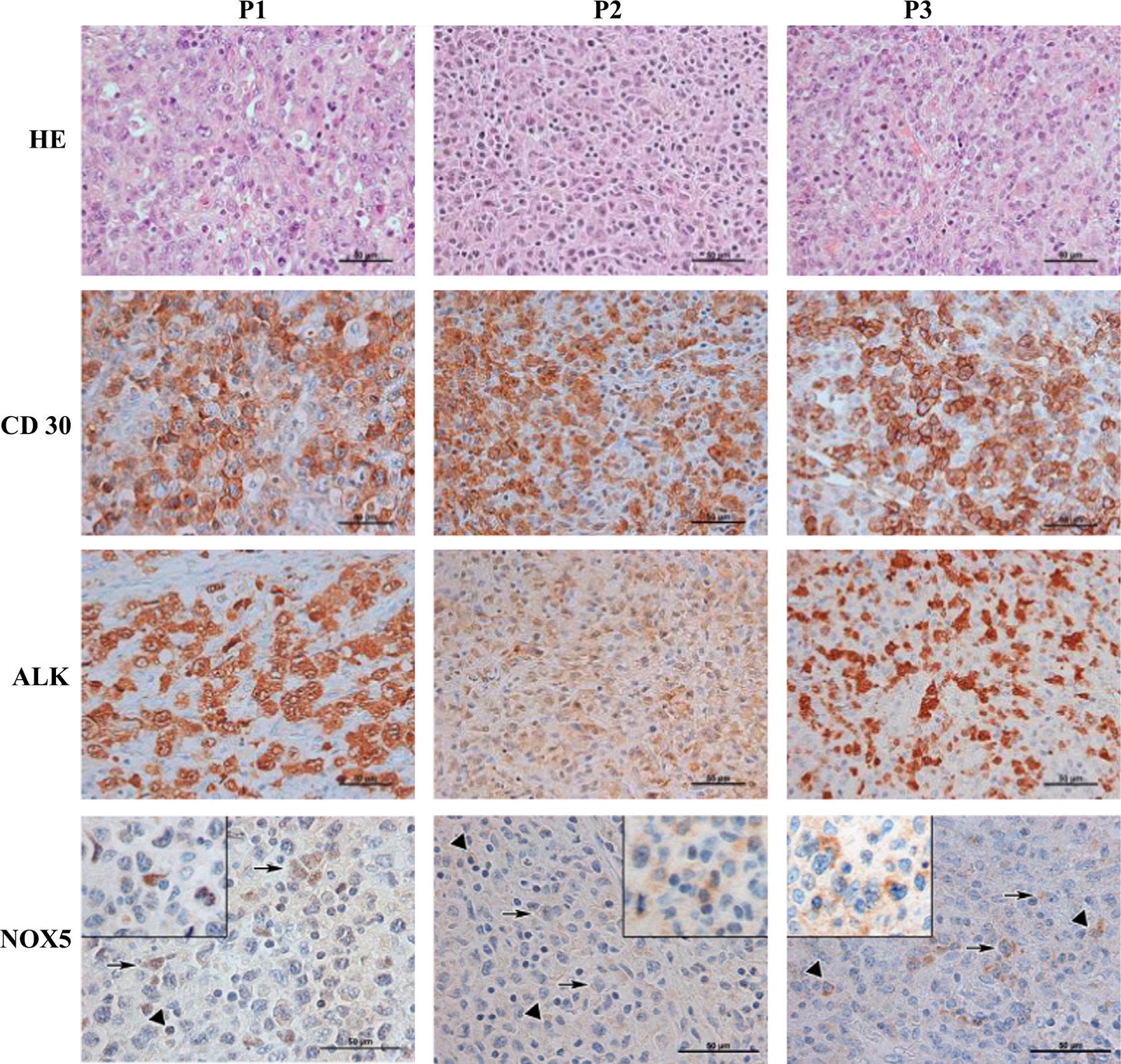
Fig. 2.

Immunohistochemical detection of NOX5 expression in pediatric ALK+ ALCL tumor samples. Three pediatric ALK+ ALCL tumoral samples were stained with hematoxylin–eosin or processed for immunohistochemical detection using CD30−, ALK−, or NOX5-specific antibodies. Triangles denote plasma cells, arrows point to tumor cells. P1, P2, P3, patient 1, patient 2, patient 3. Original magnification, 400 × (HE, CD30, and ALK) or 600 × (NOX5), with insets showing additional positive tumor cells at higher magnification.
