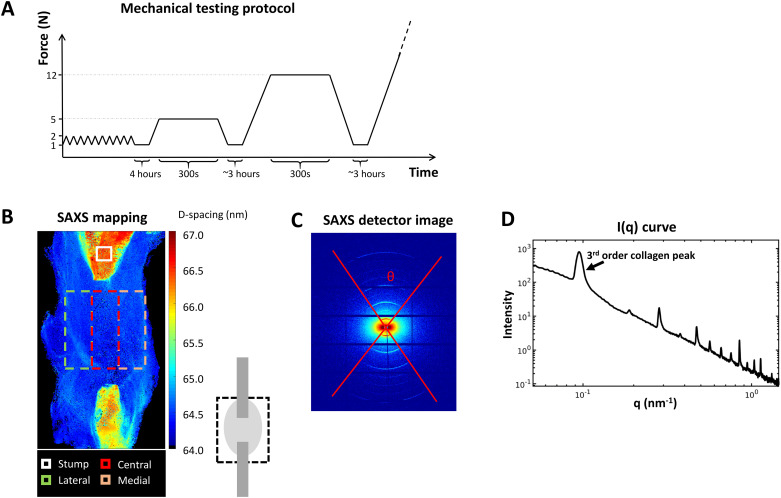
Fig 2. Analysis methods.
A) Mechanical testing protocol included preconditioning, followed by two sets of creep tests before load to failure. B) Small Angle X-ray Scattering (SAXS) mapping over the tendon callus indicates the stumps and the regions of interest for analysis in the callus. C) SAXS detector image and D) integrated scattering intensity curve obtained from (C).

