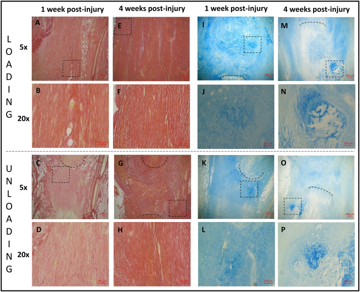
Fig 8. Histology.
Healing tendons from 1 (A, B, C, D, I, J, K, L) and 4 weeks (E, F, G, H, M, N, O, P) post-injury stained with picrosirius red (A, B, C, D, E, F, G, H) or alcain blue (I, J, K, L, M, N, O, P). Healing tendons exposed to loading (A, E, I, M (magnification 5) and B, F, J, N (magnification 20)) can be compared to healing tendons exposed to unloading with botox (C, G, K, O (magnification 5) and D, H, L, P (magnification 20)). The area of magnification 20 is shown as a square in the picture above with magnification 5 i.e. B is showing the squared area in A. The black lines are marking the tendon stump.

