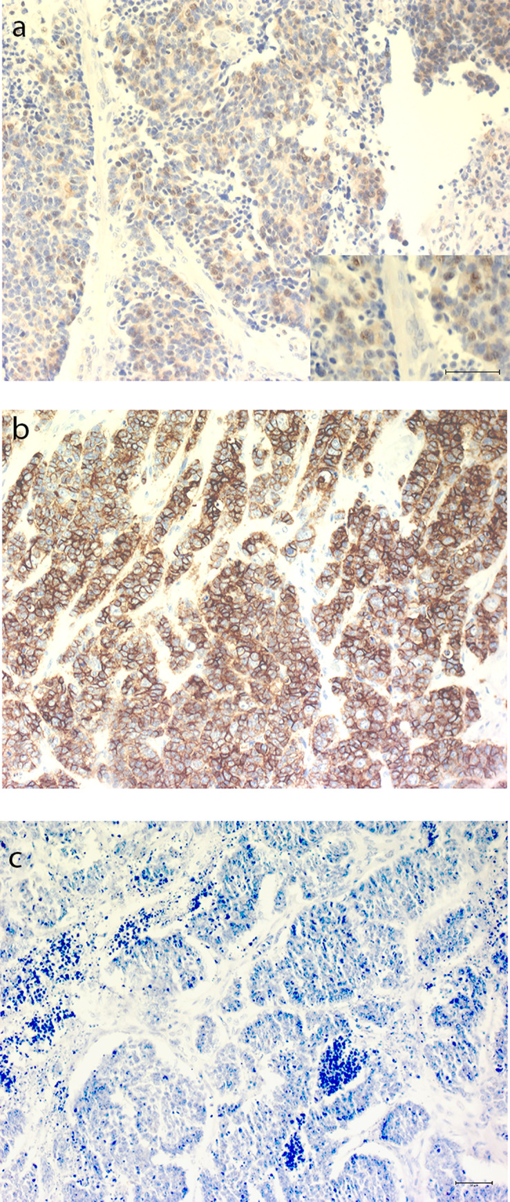
Fig 1. Representative pictures of PD-L1 immunohistochemical staining on tumors.

(a) Colon primary tumor with approximately 4% of all tumor cells immunoreactive. Insert, magnification. (b) Colon primary with 80% immunoreactive tumor cells. (c) Non-immunoreactive colon primary tumor. Scale bars 50 μm.
