Figure 3.
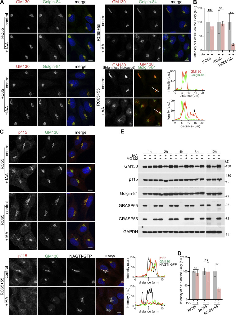
Acute simultaneous depletion of GRASP55 and 65, but not separate depletion, partially dislocates GM130 from the Golgi. (A) GM130 is partially displaced from the Golgi and relocated to the nucleus after degradation of GRASP55 together with GRASP65, but not after individual depletion of GRASP55 or 65. RC55, RC65, or RC65+55 cells were treated with doxycycline for 6 h and then with IAA for a further 2 h. The cells were then fixed and immunolabeled for GM130 (red) and the cis-Golgi protein Golgin-84 (green) and stained for DNA (blue). The brightness of the GM130 image of RC65+55 cells in the bottom panel was increased to better visualize the redistribution of GM130. The arrow indicates the nuclear signal of GM130. White lines show the position of the line-scan used to measure the fluorescence intensity of GM130 (red) and Golgin-84 (green) across the Golgi and nucleus as shown in the graphs. Scale bar, 10 µm. (B) Quantitation of the GM130 fluorescence signal on the Golgi (marked by Golgin-84) from A. n = 3 independent experiments with >50 cells analyzed per experiment and condition. ** P < 0.01; ns, not significant. Error bars represent mean ± SD. (C) Degradation of GRASP55 and 65 partially delocalizes p115 together with GM130 from the Golgi. RC55, RC65, and NAGTI-GFP–expressing RC65+55 cells were treated as in A and immunostained for p115 (red) and GM130 (green) and labeled for DNA (blue). The Golgi is marked by the GFP fluorescence signal of the Golgi enzyme NAGTI-GFP. White lines show the position of the line-scan used to measure the fluorescence intensity of p115 (red), GM130 (green), and NAGTI-GFP across the Golgi as shown in the graphs. Scale bars, 10 µm. (D) Quantitation of the p115 fluorescence signal on the Golgi marked by GM130 or NAGTI-GFP from C. n = 3 independent experiments with >50 cells analyzed per experiment and condition. ** P < 0.01; ns, not significant. Error bars represent mean ± SD. (E) GM130 is partially degraded by the proteasome after acute double depletion of GRASP55 and 65. RC65+55 cells were treated with doxycycline for 6 h, followed by IAA or IAA plus 10 µM MG132. Cell lysates were collected at the indicated time points and subjected to immunoblotting with the indicated antibodies.

