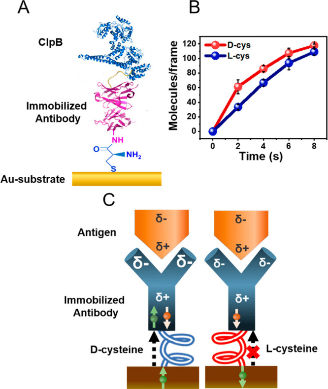
Figure 3.

Kinetics of binding of the antigen to the antibody when bound to a gold substrate through a chiral molecule. (A) Schematic showing an anti-His antibody attached to a gold substrate via either a d- or an l-cysteine linker. The surface is exposed to a solution containing the His-tagged antigen protein. (B) Number of antibody–antigen complexes as a function of time when the antibody is linked to the surface via d-cysteine (red) or l-cysteine (blue). (C) A model for the effect of the handedness of the chiral linker on the association rate (see details in text).
