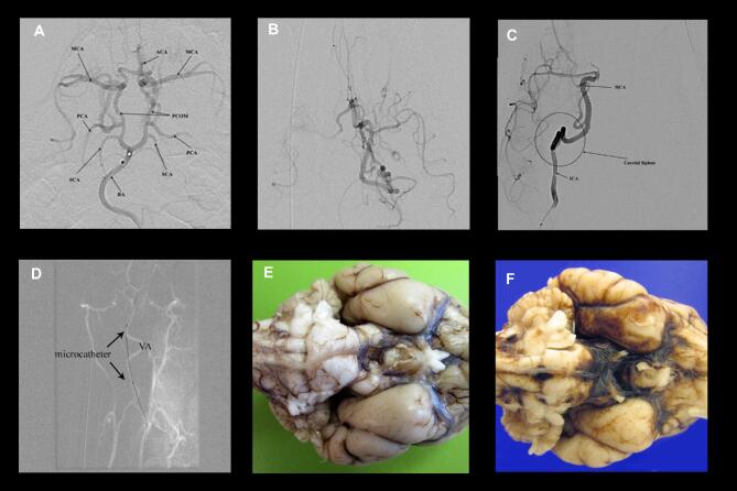
FIGURE 1.
Angiography in canine subjects. A, Diagnostic cerebral angiogram (DSA) of the circle of Willis in the canine. The microcatheter is positioned near the basilar apex (BA). Both the anterior circulation (anterior cerebral artery (ACA), middle cerebral artery (MCA)) and posterior circulation (posterior communicating artery (PCOM), posterior cerebral artery (PCA), and superior cerebellar arteries (SCAs)) are visible. B, DSA of the posterior circulation with the microcatheter positioned in the P1 PCA. Via the PCOM, the anterior circulation is partially opacified along with the PCA. This demonstrates that the canine has a posterior circulation dominance compared the anterior circulation. C, DSA of the anterior circulation with the microcatheter positioned in the proximal ICA at the bottom of the panel. The carotid siphon is very tortuous, precluding further selective access. D, microcatheter access via the vertebral artery, roadmap view. The microcatheter can be seen outside of the roadmap of the artery. This demonstrates that the vessel is highly mobile and being easily straightened by a flexible, soft microcatheter. It is likely this manipulation that led to vessel injury in posterior circulation subjects. E, Gross pathology looking at the ventral brainstem and cerebrum in subject 1, with no procedural complications. F, Same view in subject 6 demonstrating diffuse basal cistern SAH.

