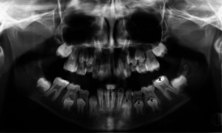
Fig. 1.
Panoramic radiograph from one of the included individuals. Teeth 31–37, which are marked with a cross, were assessed according to Demirjian et al. [15]. The criteria for the assessment of different stages varied from the beginning of calcification of crown to apical root apex completely closed (stages A–H). In Fig. 1, for example, tooth 31 was assessed as stage H and tooth 37 as stage D. If a tooth appeared to be between stages, the earlier stage was chosen

