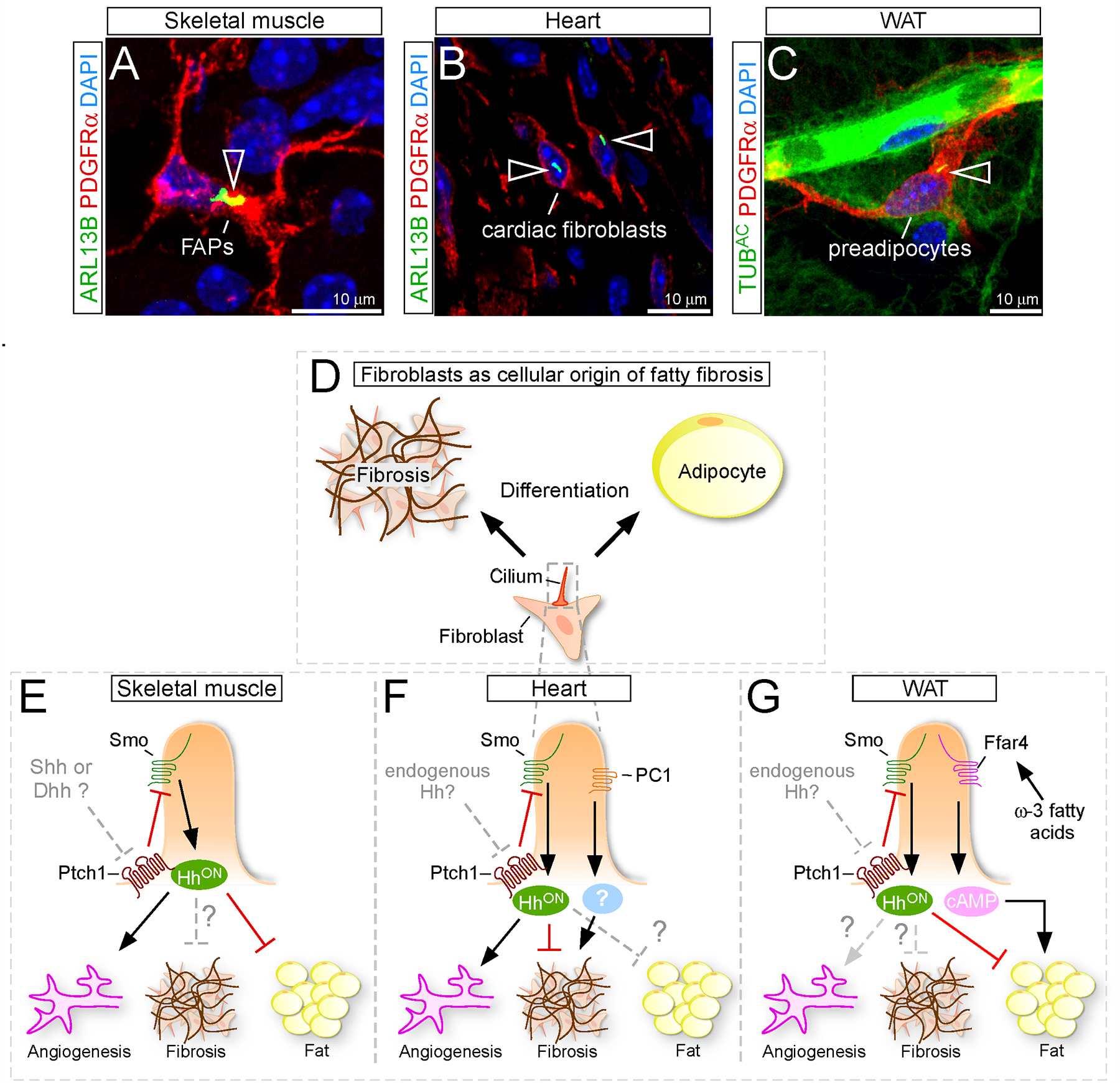
Figure 3. Ciliary control of fibroblast function during fatty fibrosis.

(A-C) Fibroblasts expressing the marker PDGFRα (red) are frequently ciliated (green; arrowhead) in different tissues such as skeletal muscle (A), heart (B) and white adipose tissue (WAT) (C). Nuclei in blue (DAPI).
(D) Fibroblasts are the cellular source of fibrotic scar and fat tissue.
(E) Ciliary Hh signaling has a pro-angiogenic and anti-adipogenic function in skeletal muscle fibroblasts. The role of Hh during fibrosis as well as which endogenous Hh ligand is being used, however, is still unclear.
(F) Hh signaling in cardiac fibroblasts (CFs) blocks fibrosis and promotes angiogenesis during ischemic injuries. It remains to be determined if Hh has an endogenous role and if Hh could also affect fat infiltration. CF cilia also utilize polycystin1 (PC1) to control fibrosis, however the exact mechanism still needs to be determined.
(G) Cilia, present on the preadipocytes in white adipose tissue (WAT), balance adipogenesis by sensing the anti-adipogenic Hh and the pro-adipogenic ω−3 fatty acid signal. If Hh has an endogenous role in WAT, however, to control angiogenesis and/or fibrosis, is unknown.
