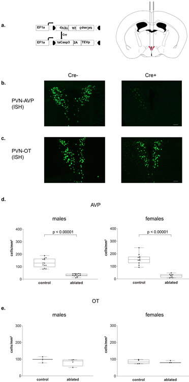
Figure 2 – Histology.
(a) Cre-dependent adeno-associated virus (AAV-flex-taCasp3-TEVp) construct and location of bilateral PVN injection site; AP −0.42 mm; ML ± 0.35 mm; DV 5.2 mm; modified from [48]. (b) Representative images of fluorescent in situ hybridization-labeled PVN AVP-expressing cells and (c) OT-expressing cells. (d) Boxplots indicating individual data points, median, first and third quartiles for PVN AVP-expressing cell number across four anterior-posterior sections in male and female subjects. Within the PVN, we found a significant decrease in AVP-expressing cell number in both Cre+ male and female subjects compared to Cre− controls (males: p < 0.00001; females: p < 0.00001). Cre− (n = 13) and Cre+ (n = 15) males and Cre− (n = 11) and Cre+ (n = 9) females. (e) We found no significant OT-expressing cell loss between Cre+ and Cre− subjects (males: p = 0.35; females: p = 0.9). Cre− (n = 3) and Cre+ (n = 4) males and Cre− (n = 4) and Cre+ (n = 3) females. Images were taken at 10x, fluorescent cells per mm2, scale bar = 50 μm.

