
Figure 1. The microperimetry test pattern used in the Fight-RP study.
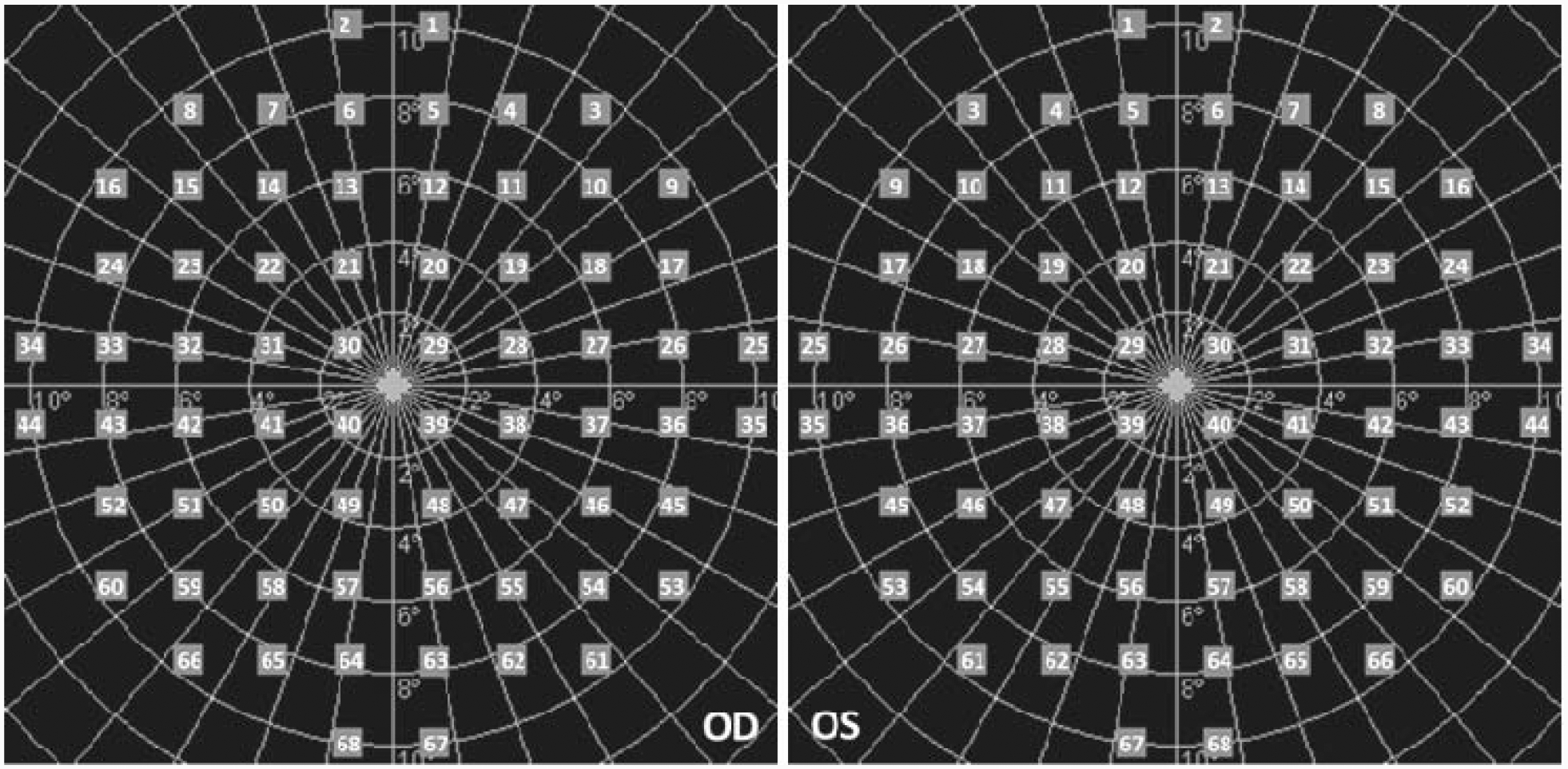
The pattern included 68 test loci covering the central 20° of the retina. Eccentricy of the test loci was categorized into 3 regions: Foveal region included 4 loci distributed on the ring of 2° eccentricity (Loci 29, 30, 39, and 40); Parafoveal region included 12 loci distributed on the ring of 4° eccentricity (Loci 19–22, 28, 31, 38, 41, 47–50); and Perifoveal region included 52 loci distributed on the rings of 6°−10° eccentricity (Loci 1–18, 23–27, 32–37, 42–46, 51–68).