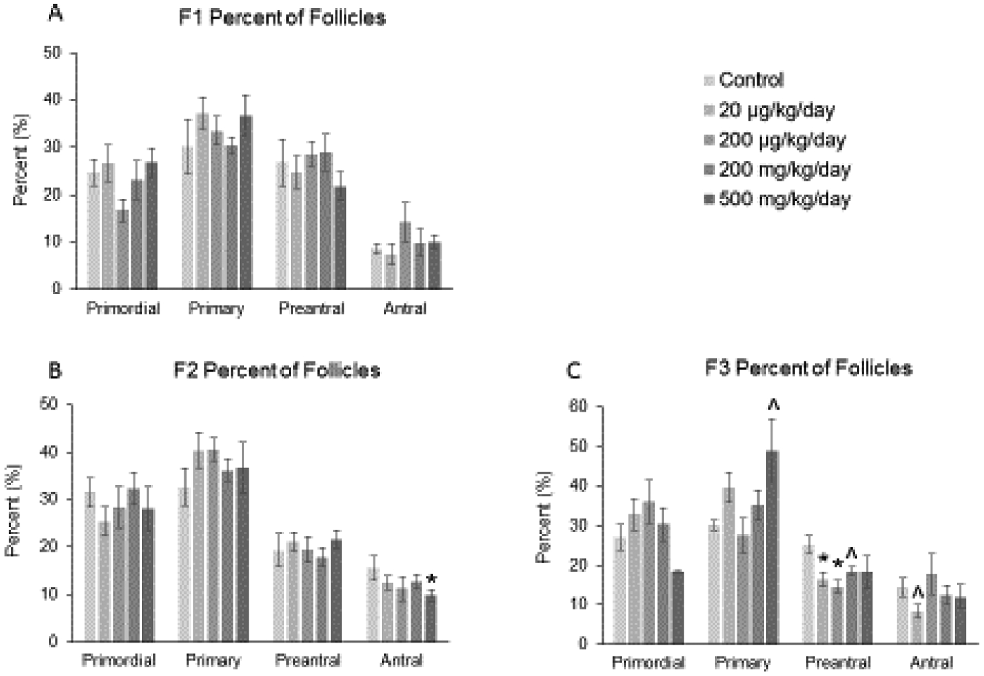
Figure 3.

Effect of prenatal exposure to the phthalate mixture on the percentage of follicle types per ovary at 13 months of age in the F1, F2, and F3 generations of mice. Ovaries from the F1, F2, and F3 generations were subjected to histological evaluation for the percentage of each follicle type. Follicles in the F1 generation (panel A; control = 5–6 females/treatment group, 20 μg/kg/day = 9 females/treatment group, 200 μg/kg/day = 9 females/treatment group, 200 mg/kg/day = 7–8 females/treatment group, 500 mg/kg/day = 9 females/treatment group), F2 generation (panel B; control = 7 females/treatment group, 20 μg/kg/day = 11 females/treatment group, 200 μg/kg/day = 7 females/treatment group, 200 mg/kg/day = 9 females/treatment group, 500 mg/kg/day = 5–6 females/treatment group), and the F3 generation (panel C; control = 5–6 females/treatment group, 20 μg/kg/day = 8–9 females/treatment group, 200 μg/kg/day = 5–6 females/treatment group, 200 mg/kg/day = 6–7 females/treatment group, 500 mg/kg/day = 3–4 females/treatment group) were counted and separated by stage of development, and percentages of each follicle type were calculated for each treatment group. Graphs represent means ± SEM in the F1 generation, F2 generation, and the F3 generation. Asterisks (*) indicate significant differences compared to the control (p < 0.05) and carets (^) indicate borderline significance compared to the control (0.05 < p < 0.1).
