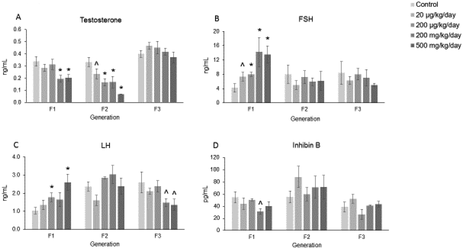
Figure 4.

Effect of prenatal exposure to the phthalate mixture on serum sex steroid, gonadotropin, and peptide hormone levels at 13 months of age in the F1, F2, and F3 generations of mice. Sera were subjected to ELISAs or RIAs for the measurements of testosterone (panel A; F1 = 7–9 females/treatment group, F2 = 5–11 females/treatment group, F3 = 4–9 females/treatment group), FSH (panel B; F1 = 5–9 females/treatment group, F2 = 6–10 females/treatment group, F3 = 4–9 females/treatment group), LH (panel C; F1 = 6–9 females/treatment group, F2 = 6–11 females/treatment group, F3 = 4–9 females/treatment group), and inhibin B (panel D; F1 = 7–9 females/treatment group, F2 = 6–10 females/treatment group, F3 = 4–9 females/treatment group). Graphs represent means ± SEM in the F1 generation, F2 generation, and the F3 generation. Asterisks (*) indicate significant differences compared to the control (p < 0.05) and carets (^) indicate borderline significance compared to the control (0.05 < p < 0.1).
