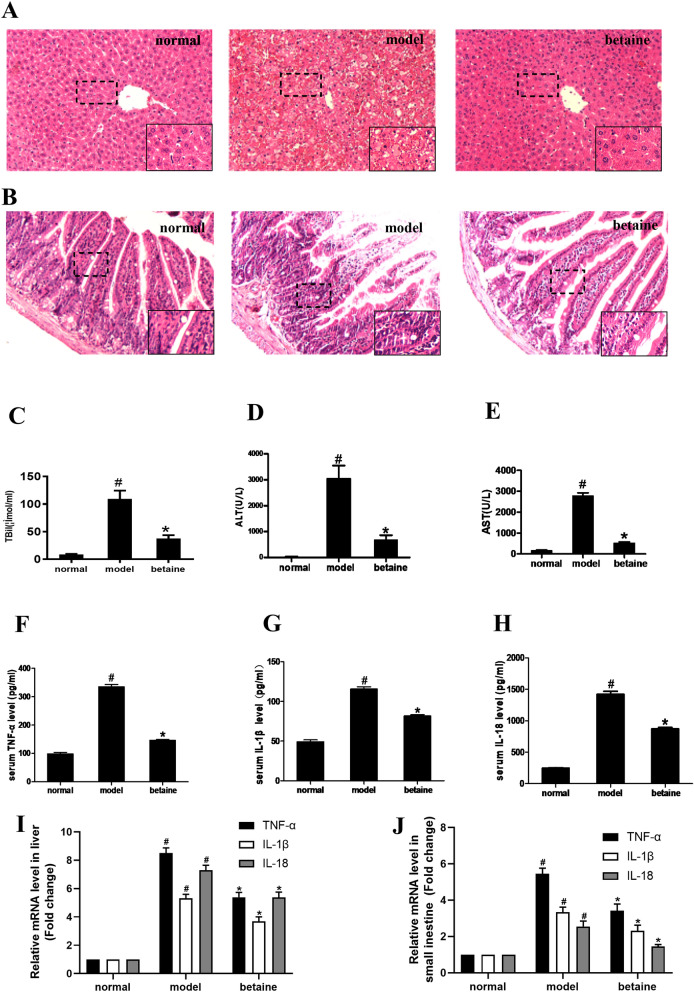
Figure 1.
Effect of betaine on liver and small intestine tissue pathological changes and serum biochemical indicators in ALF mice. (A) The liver tissues were stained with HE (× 200). (B) The small intestine tissues were stained with HE (× 200). (C–E) The serum levels of ALT, AST, and TBIL in different animal groups. #P < 0.05, compared with the normal group; *P < 0.05, compared with the model group. (F–H) The serum levels of TNF-α, IL-1β and IL-18 in each group. (I,J) The relative mRNA levels of TNF-α, IL-1β and IL-18 in liver and small intestine tissue. #P < 0.05, compared with the normal group; *P < 0.05, compared with the model group.

