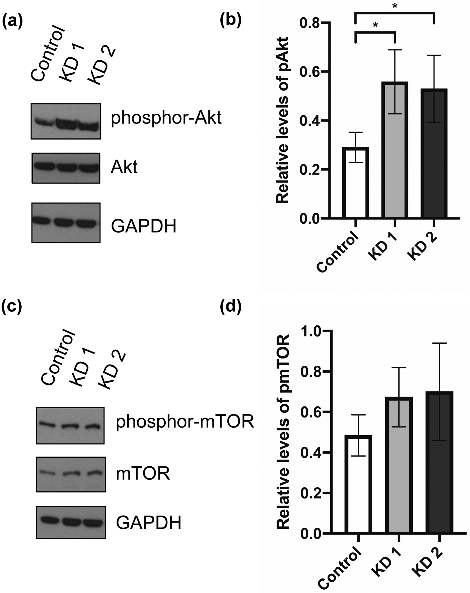
Figure 4. SST knockdown increases phosphorylation of Akt.

Representative Western blots (repeated 3 or more times) of SST-knockdown and control BON cells. Protein was collected at day 5 after siRNA transfection for Western blot analysis. (a) Blots were incubated with antibodies against phosphor-Akt (Ser473), total Akt, and GAPDH. (b) Relative levels of phosphor-Akt, normalized to GAPDH, between SST-knockdown and control cells. (c) Blots were incubated with antibodies against phosphor-mTOR (Ser2448), total mTOR, and GAPDH. (c) Relative levels of phosphor-mTOR, normalized to GAPDH, between SST-knockdown and control cells. Quantification performed by ImageJ analysis. Data represent mean ± SD and are representative of 3 or more experiments. * p < .05.
Abbreviations: short interfering RNA (siRNA), knockdown (KD), somatostatin (SST)
