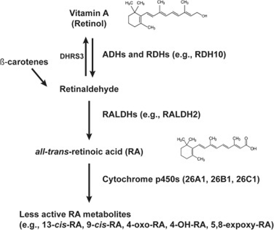
Figure 1. Retinoic acid metabolism.

The availability of RA is tightly controlled in the mammalian CNS. Mammals are unable to synthesize RA de novo and require intake of vitamin A or other precursors (β-carotenes) from food sources. Vitamin A is converted to retinaldehyde by alcohol dehydrogenases (ADHs) and retinol dehydrogenases (RDHs). In mouse, RDH10 is necessary for conversion of retinol to retinaldehyde in the developing embryo [38]. Enzymes such as Short-chain Dehydrogenase/Reductase 3 (DHRS3) facilitates the reverse transformation of retinaldehyde to retinol [40, 68]. Retinaldehyde is further oxidized to form RA by aldehyde dehydrogenases (ALDHs or RALDHs) in an irreversible step. RALDH2 is critical for RA synthesis during early CNS development [42]. Cytochrome p450 26 subfamily enzymes regulate RA levels in the embryo and catalyze reactions to reduce RA bioavailability by converting RA to 4-OH-RA, 4-oxo RA, and other oxidized, less active metabolites [43]. These metabolites undergo glucuronidation which promote elimination pathways [180].
