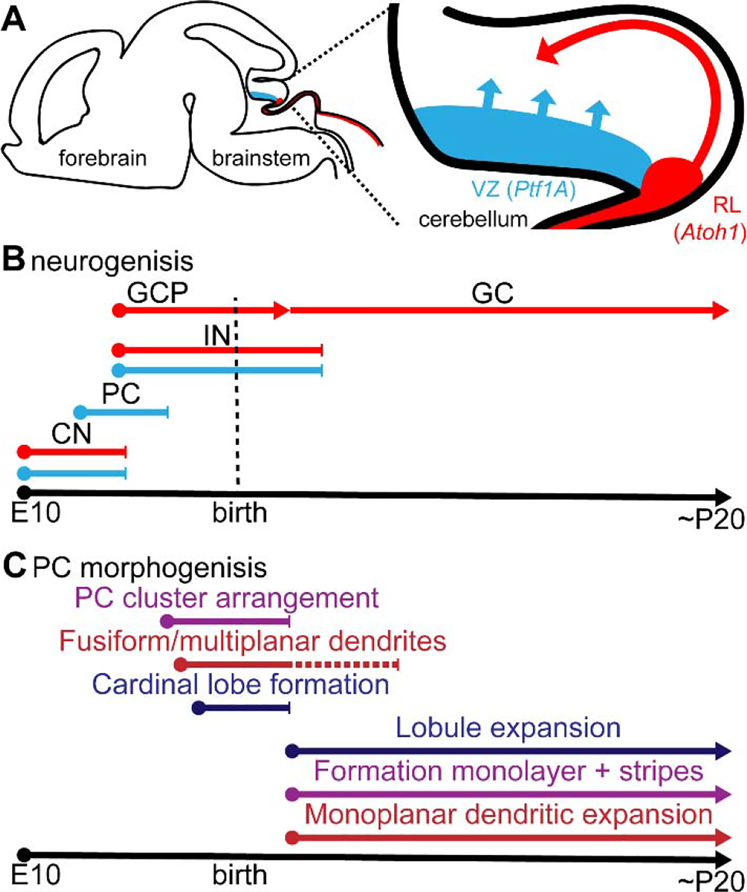
Figure 2. Cerebellar cell-type identities are spatially and temporally determined in the embryo according to birthplace and birthdate.

A. Schematic of a sagittal section of an embryonic mouse brain (left). The inset (right) is zoomed in onto the cerebellar anlage. The ventricular zone (VZ) is shown in blue, progenitor cells in this region express the transcription factor Ptf1a and migrate radially into the core of the cerebellar anlage after final mitosis. The rhombic lip (RL) is shown in red, progenitor cells in this region express the transcription factor Atoh1 and migrate over the cerebellar surface in a tangential manner. B. Neurogenesis of diverse cerebellar cell types occurs in a temporally controlled manner. Blue lines indicate neurons developing from the VZ, red lines indicate neurons developing from the RL. CN = cerebellar nuclei; PC = Purkinje cell; IN = interneurons; GCP = granule cell precursor; GC = granule cell. C. Purkinje cell morphogenesis occurs in two distinct phases (the panel is split into processes, at top and bottom of the timeline). The processes shown at the bottom are dependent on granule cells. Purple lines indicate temporally distinct migratory paths. Red lines indicate the morphogenesis of the Purkinje cell dendritic arbor. Blue lines indicate lobule formation.
