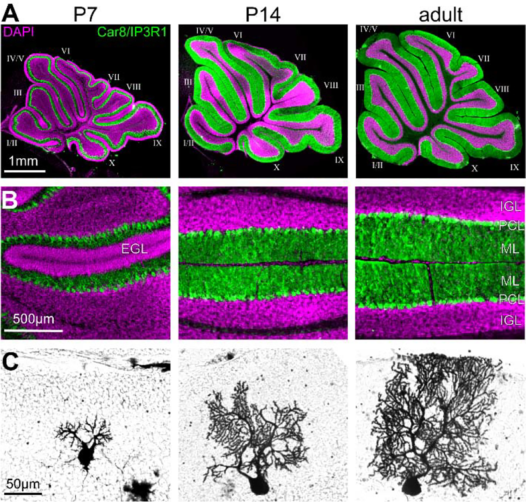
Figure 4. Expansion of the cerebellar cortex in the anterior-posterior axis during development.

A. Sections cut through the cerebellar vermis of P7, P14, and adult mice stained for the nuclear marker DAPI and the Purkinje cell markers, Car8 and IP3R1. B. High-power images of the cerebellar cortex stained for the nuclear marker DAPI and the Purkinje cell markers, Car8 and IP3R1. Note the large external granular cell layer (EGL) in P7 animals and the increase in the thickness of the molecular layer (ML) between the different time points. C. Example of Golgi-Cox-stained Purkinje cells at P7, P14, and adult.
