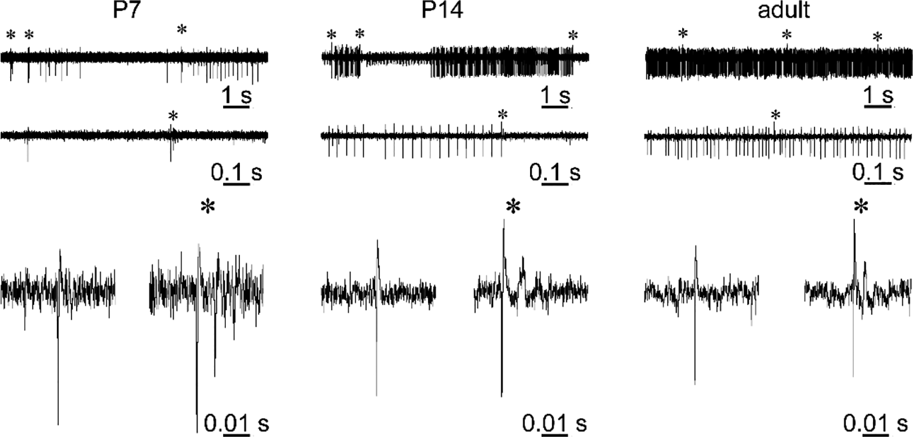
Figure 5. The electrophysiological signature of Purkinje cells changes during development.

Example traces of in vivo extracellular electrophysiology recordings from anesthetized P7, P14, and adult mice. Top traces are 10 seconds of recordings, middle traces are 1 second of recording, and bottom traces are examples of simple spikes (left) and complex spikes (right) from each different age. Complex spikes are indicated with an asterisk (*). During development, Purkinje cells start to fire more frequently and more regularly with less pauses between simple spike trains.
