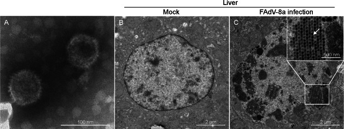
Fig. 7.
Electron micrographs of FAdV-8a. A Electron micrographs of FAdV-8a from LMH cells. Typical icosahedral and non-enveloped particles (70 nm in diameter) were observed. B Electron micrographs of a control chicken liver at 4 dpi. C Electron micrographs of FAdV-8a (indicated by arrows) on a FAdV-8a-challenged chicken liver at 4 dpi.

