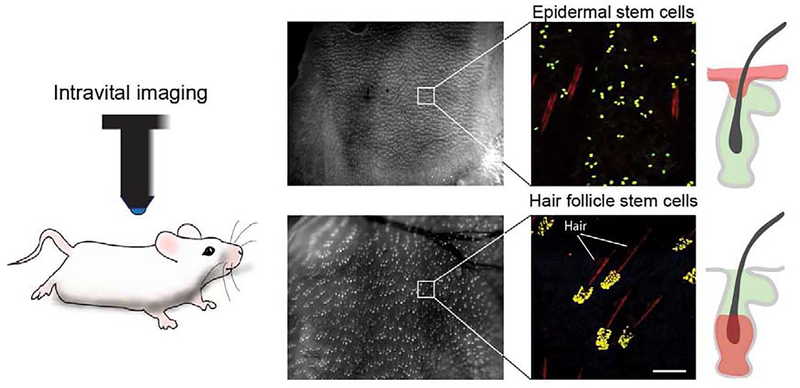
Figure 1. Genetic labeling and visualization of single keratinocytes in live mouse skin.
Representative low and high magnification images demonstrating high-throughput visualization of single-labeled stem cells in the mouse skin, by intravital imaging. Cells in the epidermis and hair follicles can be differentially labeled using cell-type specific inducible drivers. Scale bar 100μm.

