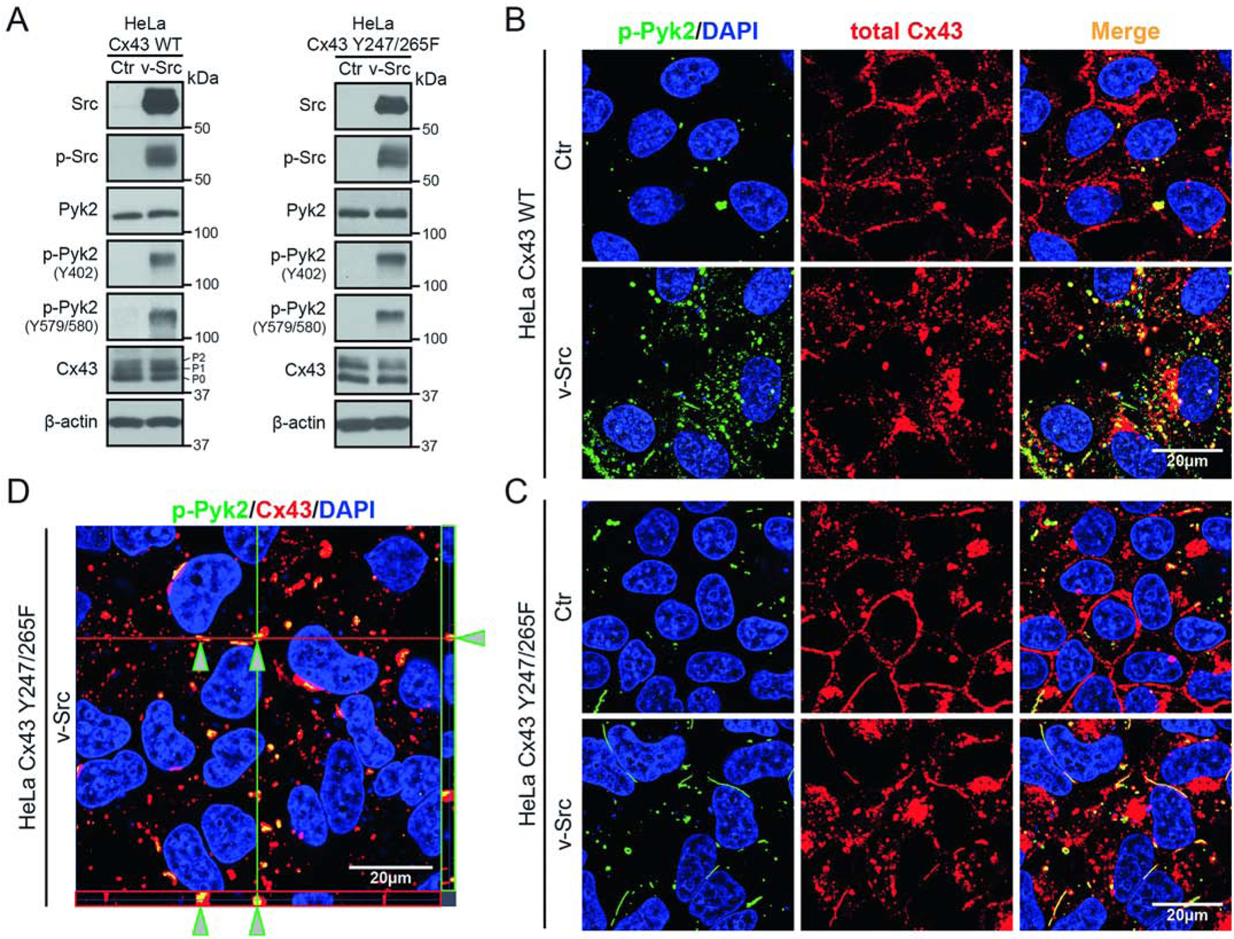
Figure 1: Active Pyk2 interacts with Cx43 in HeLa cells.

(A) Western blot of lysate from HeLa cells stably expressing Cx43 WT or Cx43 Y247/265F ± v-Src (24 h). Antibodies used are labeled on the left of each panel. The Cx43 P0, P1, and P2 isoforms have been labeled. Cellular localization of (B) Cx43 WT or (C) Cx43 Y247/265F ± v-Src (24 h) in HeLa cells detected by immunofluorescence (green, p-Pyk2Y579/580; blue, DAPI-stained DNA; red, total Cx43; yellow, p-Pyk2/Cx43 colocalization). (D) Z-stack imaging of HeLa Cx43 Y247/265F cells after v-Src transfection. Arrows point to Cx43 colocalization with active Pyk2.
