
Figure 2: Inhibition of active Pyk2 decreases Cx43 phosphorylation at residues Y247, Y265, and Y313.
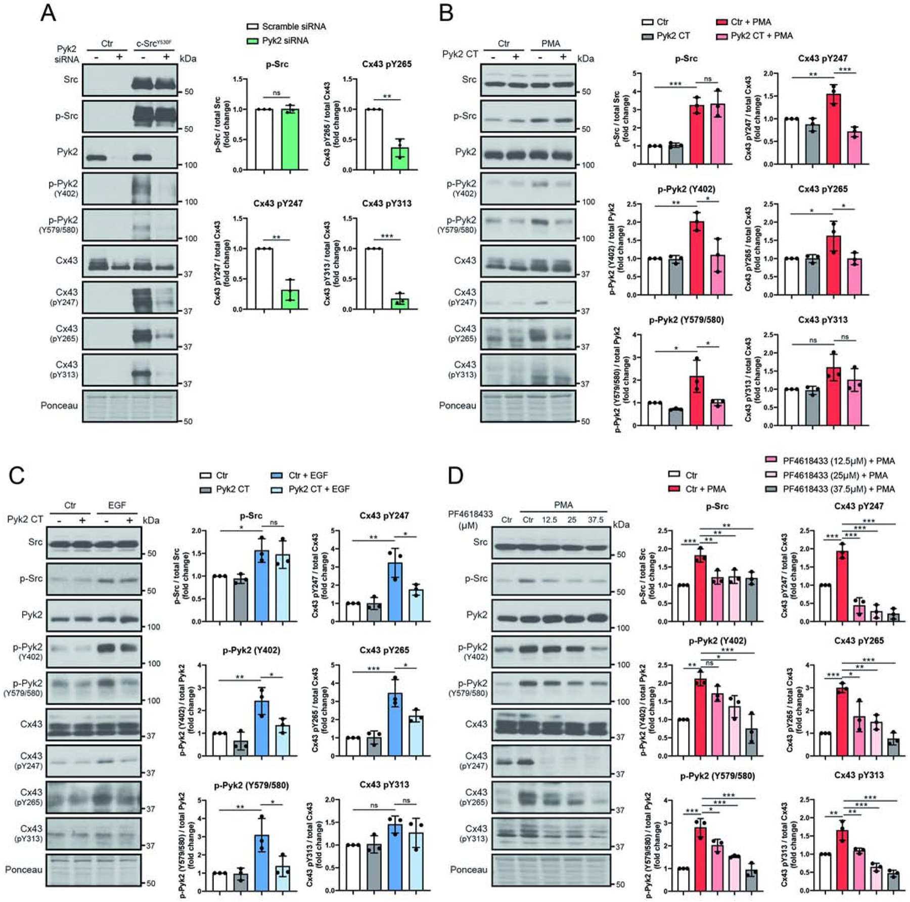
Lysate from HeLaCx43 cells treated with (A) Pyk2 siRNA and c-SrcY530F (active c-Src, 24 h), (B) Pyk2 CT and PMA (100 nM, 1 hr), (C) Pyk2 CT and EGF (100 ng/mL 1 hr), or (D) Pyk2 inhibitor PF4618433 and PMA (100 nM, 1 hr) were Western blotted. Antibodies used are labeled on the left of each panel. Protein levels were quantified by analyzing scanned blots using ImageJ software, with normalization of protein expression to the control lane (value set arbitrarily as 1). Phosphorylated protein levels were normalized to total protein. Data are representative of three independent experiments (one-way ANOVA, *P<0.05, **P<0.01, ***P<0.001).