
Figure 5: PMA mediated Cx43 gap junction closure is reversed by inhibiting active Pyk2 and c-Src.
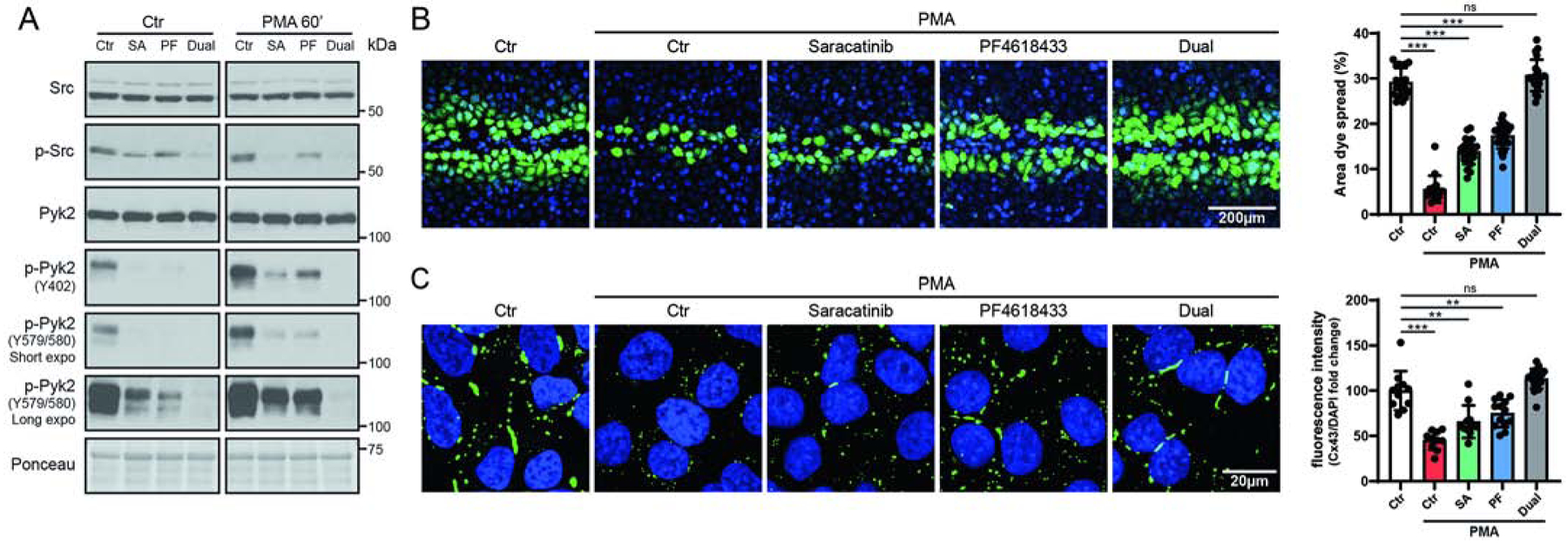
(A) Lysate from PMA treated (100 nM, 60 min) HeLaCx43 ± Src (Saracatinib, SA, 5μM) and/or Pyk2 (PF4619433, PF, 50μM) inhibitors was Western blotted. Antibodies used are labeled on the left of each panel. (B) Level of gap junction intercellular communication in HeLaCx43 cells pre-treated with Saracatinib, PF4618433 or both and then treated with PMA (60 min) was determined using the scrape loading dye transfer assay. Provided are representative immunofluorescence images (green, Lucifer yellow; blue, DAPI). Quantification of the area of Lucifer Yellow transfer shows the effect of inhibiting Src and/or Pyk2 phosphorylation of Cx43. Immunofluorescence spread (area) of Lucifer Yellow transfer was measured by ImageJ software. (C) Representative fluorescent images and quantification of in situ TX-100 extracted Cx43 (green, Cx43; blue, DAPI). Quantification was determined by normalizing the mean fluorescent intensity of insoluble Cx43 to DAPI. All data are representative of three independent experiments (one-way ANOVA, **P<0.01, ***P<0.001).