Figure 3.
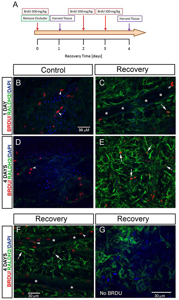
Proliferation of choroidal cells during recovery from myopia. (A) Scheme of the BrdU labeling experiment. BrdU was injected intraperitoneally into chicks following 10 days of form deprivation (day 0 recovery), followed by immediate removal of occluders to induce recovery. Additional BrdU injections were administered following 2 and 3 days of recovery. Tissue was harvested following 1 and 4 days of recovery (n = 3 chicks/ time point). After isolation of choroids, RALDH2 and BrdU immunolabeling was performed simultaneously as described in Materials and Methods and imaged using confocal microscopy. (B- F) Representative merged confocal images demonstrating RALDH2 and BrdU immunopositive cells. Intensely labelled RALDH2 positive cells (Alexa 488-labelled, green) were detected in choroids following 1 and 4 days of recovery (C,E,F,G). Proliferating RALDH2 positive cells were identified by the presence of BrdU labeling (Alexa 568-labelled, red) in the nucleus, some of which varied in intensity (C,E,F, arrows). Additional proliferating choroidal cells (RALDH2-negative) were identified by nuclear BrdU labelling (B,C,D, F, arrowheads). (G) No alexa-568 fluorescence was detected in tissues from chicks that were not administered BrdU.

