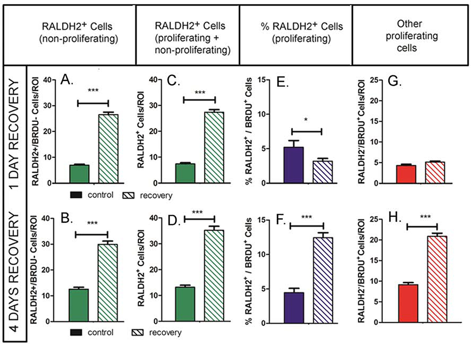
Figure 4.
Quantitation of cell proliferation during recovery from myopia. (A, B) The average number of nonproliferating RALDH2 positive cells (RALDH2+/BRDU−) was significantly higher in recovering eyes following 1 and 4 days of recovery. (C, D) The total number of proliferating and non-proliferating RALDH2 positive cells (RALDH2+/BRDU− and RALDH2+/BRDU+) was significantly higher in recovering eyes following 1 and 4 days of recovery. (E, F) The percentage of choroidal RALDH2 positive cells that were proliferating (%RALDH2+/BRDU+) was significantly decreased following 1 day of recovery compared to contralateral controls, but was significantly increased in recovering eyes after 4 days of recovery. (G, H) The number of proliferating choroidal cells that were RALDH2 negative (RALDH2-/BrdU+) was significantly increased following 4 days of recovery. Results were calculated as the average of the total number of RALDH2-positive cells per each 212 μm x 212 μm x 1 μm (x, y, z, respectively) slice within each region of interest (ROI) (n = 2 – 4 ROI’s for n = 3 separate choroids/condition). Solid bars represent control choroids; hashed bars represent recovering choroids in A – H. 2 – 4 separate pixel areas were evaluated in 3 separate choroids from control and treated eyes at each time point. ***p < 0.001; *p < 0.05 (Student’s t-test).

