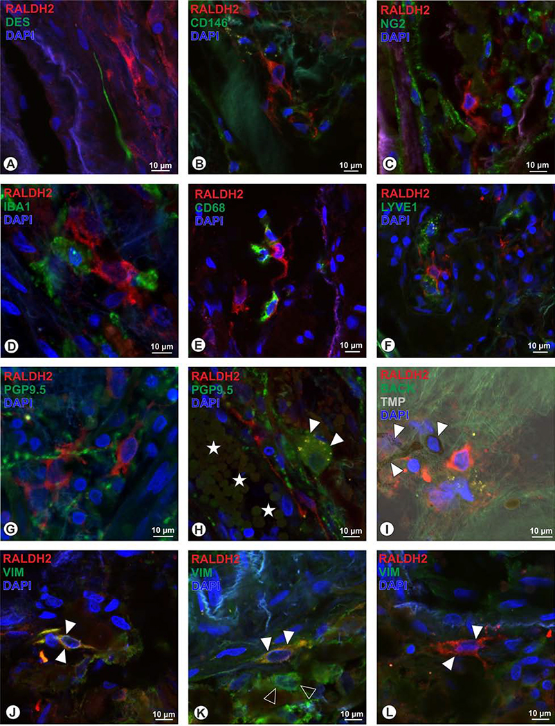
Figure 8.
Human RALDH2+ cells (red) could not be identified as pericytes, as the pericyte markers desmin (A, green), CD146 (B), and NG2 (C), revealed no co-localization. Further, the microglia marker IBA1 (D), and the macrophage markers CD68 (E) and LYVE1 (F) were absent in RALDH2+ cells. RALDH2+ cells do not belong to neuronal cell populations since PGP9.5 (G) revelaed no overlap, and further intrinsic choroidal neurons were lacking RALDH2 (H, arrowheads; yellow dots within the neuron represent lipofuscin granules; asterisks: choroidal blood vessel). When applying the trans-ilumination mode in the confocal microscope (TMP, I), melanocytes (arrowheads) could be identified by the presence of melanin granules, but were not co-localized for RALDH2. J to L: the intermediate filament vimentin (VIM) was co-localized in some RALDH2+ cells (arrowheads in J, K), while also other cells were detected, that displayed VIM only (open arrowheads, K) and further a subpopulation of RALDH2+ cells were lacking VIM immunoreactivity (arrowheads in L).

