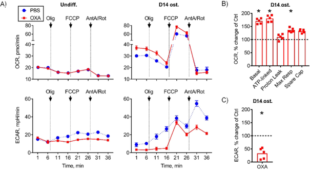
Figure 2. LDH inhibitor oxamate diverts cell bioenergetics from glycolysis to oxidative phosphorylation.
Bioenergetic profiling of undifferentiated and osteoinduced C3H10T1/2 cells in the presence of 1 mM OXA or PBS using Seahorse XF96. Oxygen consumption rate (OCR) measures OxPhos activity and extracellular acidification rate (ECAR) measures glycolytic activity. (A) OCR and ECAR measurements were taken at baseline and after addition of Olig (Oligomycin A, inhibitor of mitochondrial ATP-Synthase), FCCP (Carbonyl cyanide-p-trifluoromethoxyphenylhydrazone, protonophore which uncouples mitochondrial proton gradient), and AntA/Rot (Antimycin A and Rotenone, inhibitors of mitochondrial electron transport chain complexes III and I respectively). Undifferentiated cells have minimal reliance on OxPhos and treatment with OXA had no effect on OxPhos or basal glycolytic activity (left panel). In osteoinduced C3H10T1/2 cells, OXA treatment led to inhibition of glycolysis and further activation of OxPhos (right panel). Osteoinduced C3H10T1/2 cells treated with OXA had (B) significantly increased OCR values, including basal, ATP-linked, and maximal respiration, and (C) significantly reduced ECAR values. This demonstrates that OXA stimulates OxPhos and inhibits glycolysis in osteoinduced cells. Data are presented as boxplots with actual data points, median, and range. P value was determined by unpaired t-test and shown in data sets with p < 0.05.

