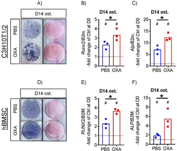
Figure 3. LDH inhibition increases osteogenic potential of both mouse and human osteoprogenitors.
C3H10T1/2 cells osteoinduced for 14 days in the presence of 1 mM OXA show increased ALP (A, left column) and Alizarin Red (A, right column) staining and expression of osteogenic markers Runx2 (B) and Alp (C). Human BMSCs osteoinduced for 14 days in the presence of 1 mM OXA show higher levels of ALP (A, left column) and Alizarin Red (A, right column) staining and RUNX2 (E) and ALP (F) expression. Images are representative of 6 (3 independent batches of cells, 2 technical replicates). Data are presented as boxplots with actual data points, median, and range. P value of D14 vs D0 (vertical text) or OXA vs PBS (horizontal text) was determined by unpaired t-test and shown in data sets with p < 0.05.

