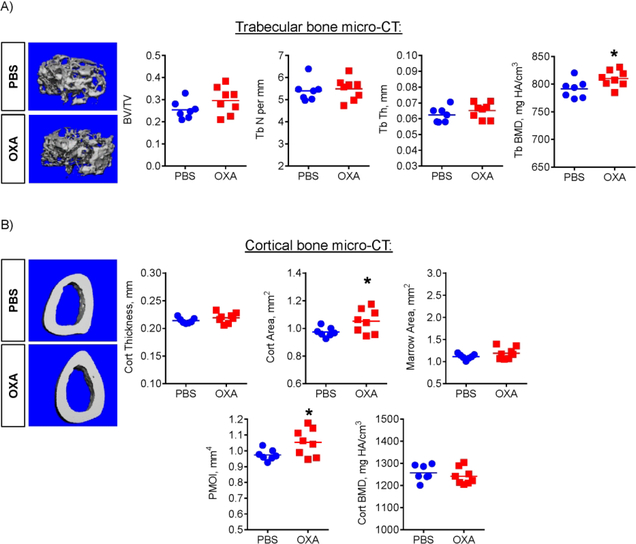
Figure 5. LDH inhibition improves trabecular bone density and cortical bone architecture in mice.
Effect of OXA on femur bone architecture, as analyzed by micro-CT. OXA-treated mice showed increased (A) trabecular bone mineral density (BMD) and (B) cortical area and polar moment of inertia (PMOI). Data are presented as boxplots with actual data points, median, and range. P value was determined by unpaired t-test and shown in data sets with p < 0.05.

