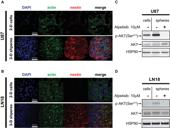
Figure 1.
Increased PI3K/AKT activation in nestin expressing 3-D spheroids. (A, B) U87 (A) and LN18 (B) cells were grown in 2-D adherent cell culture (top panels) or as 3-D spheroids in cancer stem cell medium (bottom panels) and stained for DNA (blue), actin (green) or nestin (red). Corresponding 2-D and 3-D confocal microscopy images were acquired using identical settings. Scale bar, 50 µm. (C, D) U87 (C) or LN18 (D) cells were grown as 2-D monolayer (cells) or 3-D spheroids (spheres), treated with alpelisib (10 µM, 90 min), and subjected to immunoblotting using rabbit anti p-AKT(Ser473) or HSP90 or mouse anti AKT antibodies simultaneously, followed by detection using anti-rabbit HRP or anti-mouse AlexaFlour488 secondary antibodies.

