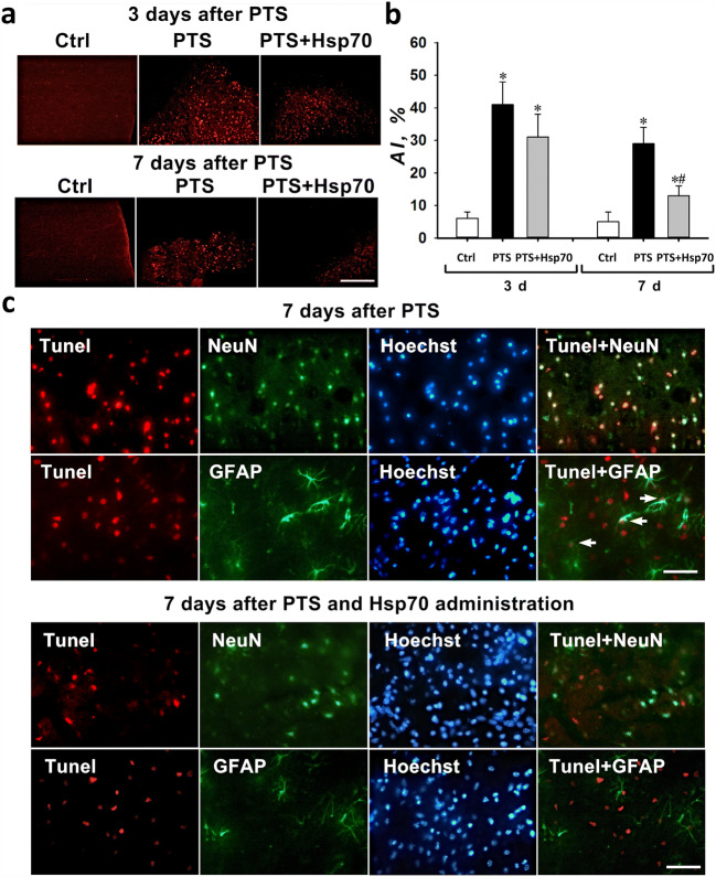
Fig. 1.
Apoptosis of penumbra cells in the cortex of mice after photothrombotic stroke. a Typical fluorescence images of the Tunel-stained (red) cortex of mice at 3 and 7 days after a photothrombotic stroke and the administration of eHsp70. Scale bar 100 μm. b The changes of the apoptotic coefficient (AI, %) in different mice groups: sham-operated control mice (Ctrl) that were intranasally injected with a physiological solution; and 3 or 7 days after photothrombotic stroke in animals that were administered intranasally with a physiological solution or 3 and 7 days after PTS and on the background of the eHsp70 introduction 3 and 7 days after PTS (after injection of Hsp70 dissolved in physiological saline). n = 24 (three fields of view were analyzed for each mouse in groups consisting from eight animals). M ± SEM. One-way ANOVA. *P < 0.05 relative to the group of sham-operated animals, #P < 0.05 relative to the group with PTS. c Colocalization of Tunel-positive nuclei (red) and neuron marker NeuN (green) or astrocyte marker GFAP (green) in penumbra 7 days after a photothrombotic stroke. Arrows indicate astrocytes with apoptotic nuclei. Scale bar 100 μm

