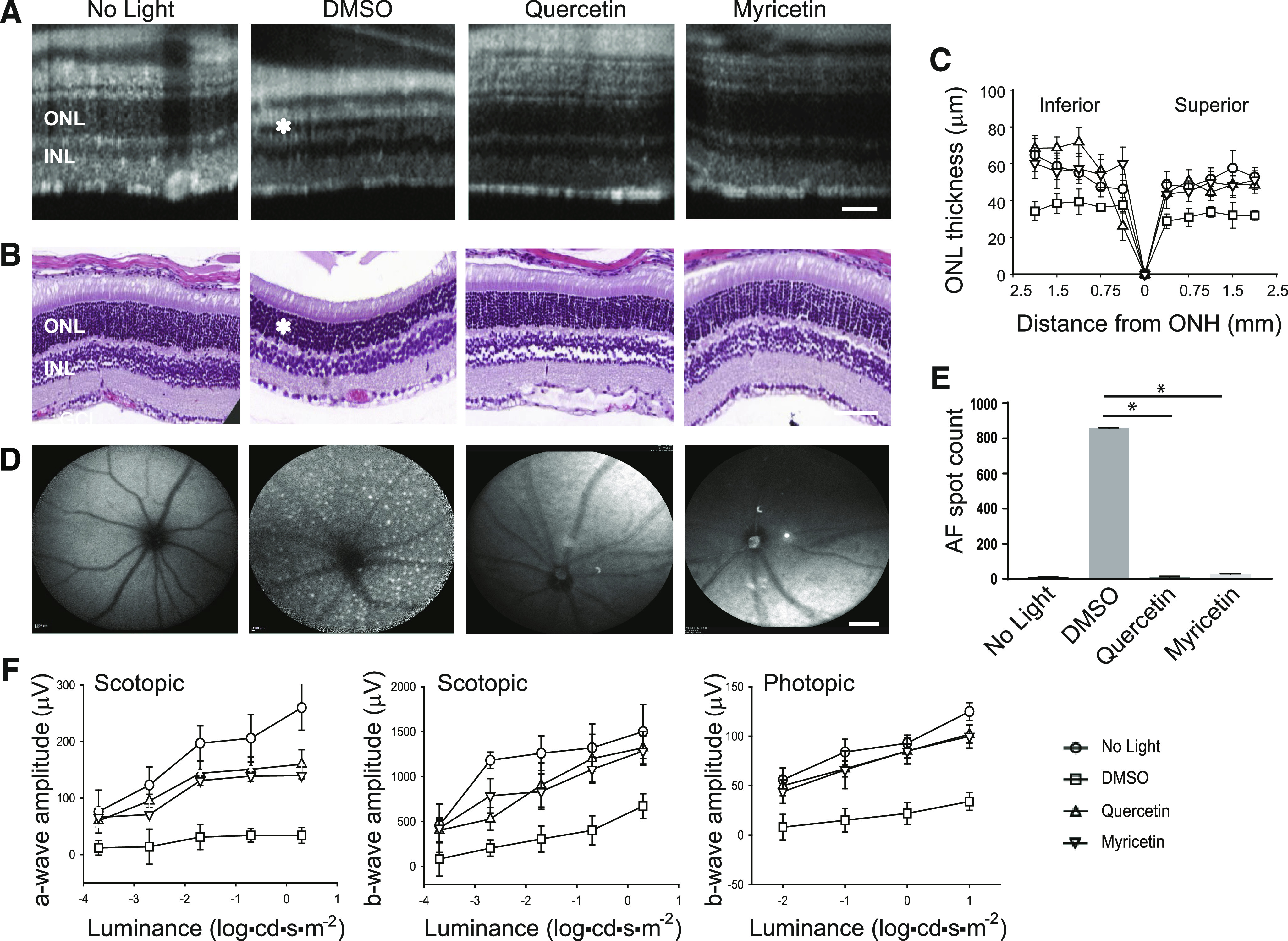
Fig. 2.

The effect of flavonoids on bright light–induced retinal degeneration in WT mice. Flavonoids were intraperitoneally injected into BALB/c mice (20 mg/kg b.wt.) 30 minutes before exposure to light at 12,000 lux for 2 hours. Then mice were kept in the dark for 7–10 days before examination of retinal morphology and function. (A) Representative OCT images. The ONL was protected in mice treated with either quercetin or myricetin as compared with DMSO-treated control mice. Asterisk indicates a shorter photoreceptor layer in DMSO-treated control mice. Scale bar, 50 µm. (B) Examination of retinal morphology after H&E staining of paraffin sections of eyes collected from mice either kept in the dark or exposed to light after indicated treatment. Asterisk indicates a shorter photoreceptor layer in DMSO-treated control mice. Scale bar, 50 µm. (C) The ONL thickness measured at inferior and superior sites at 250, 500, 750, 1000, 1500, and 2000 μm from the ONH in mice kept in the dark and mice treated with DMSO vehicle or flavonoid before exposure to bright light. Measurements were performed in 20 mice, with five mice per treatment group. Error bars indicate S.D. Changes in the ONL thickness observed between dark-adapted group and DMSO-treated, exposed-to-light group were statistically different. Changes in the ONL thickness observed after treatment with flavonoids compared with the DMSO-treated group were statistically different. No significant difference in the ONL thickness was observed between mice kept in the dark and those treated with flavonoids. (D) Representative SLO images. AF spots were detected in the retina of mice treated with DMSO before exposure to bright light. Only a few AF spots were found in the retina of mice treated with flavonoid before illumination and in mice kept in the dark. Scale bar, 50 µm. (E) Quantification of AF spots was performed in 20 mice, with five mice per treatment group. Error bars indicate S.D. Changes in the number of AF spots compared between dark-adapted group and DMSO-treated, exposed-to-light group were statistically different. Changes in the number of AF spots after treatment with either quercetin or myricetin compared with the DMSO-treated group were statistically different and are indicated with (*). No significant difference was observed between mice unexposed to light and those treated with flavonoids. (F) Retinal function examined by ERG responses. Retinal function was significantly protected in mice treated with flavonoid before exposure to bright-light insult as compared with DMSO-treated mice in both scotopic a- and b-waves and in photopic b-waves. ERG measurements were carried out in 20 mice, with five mice per treatment group. Changes in the ERG responses compared between dark-adapted mice and DMSO-treated, exposed-to-light mice were statistically different. Changes in the ERG responses after treatment with flavonoids compared with DMSO-treated mice were statistically different. No statistical difference was observed between the dark-adapted group and mice treated with flavonoids. Statistical analysis was performed with the one-way ANOVA and post hoc Dunnett’s tests.
