
Fig. 7.
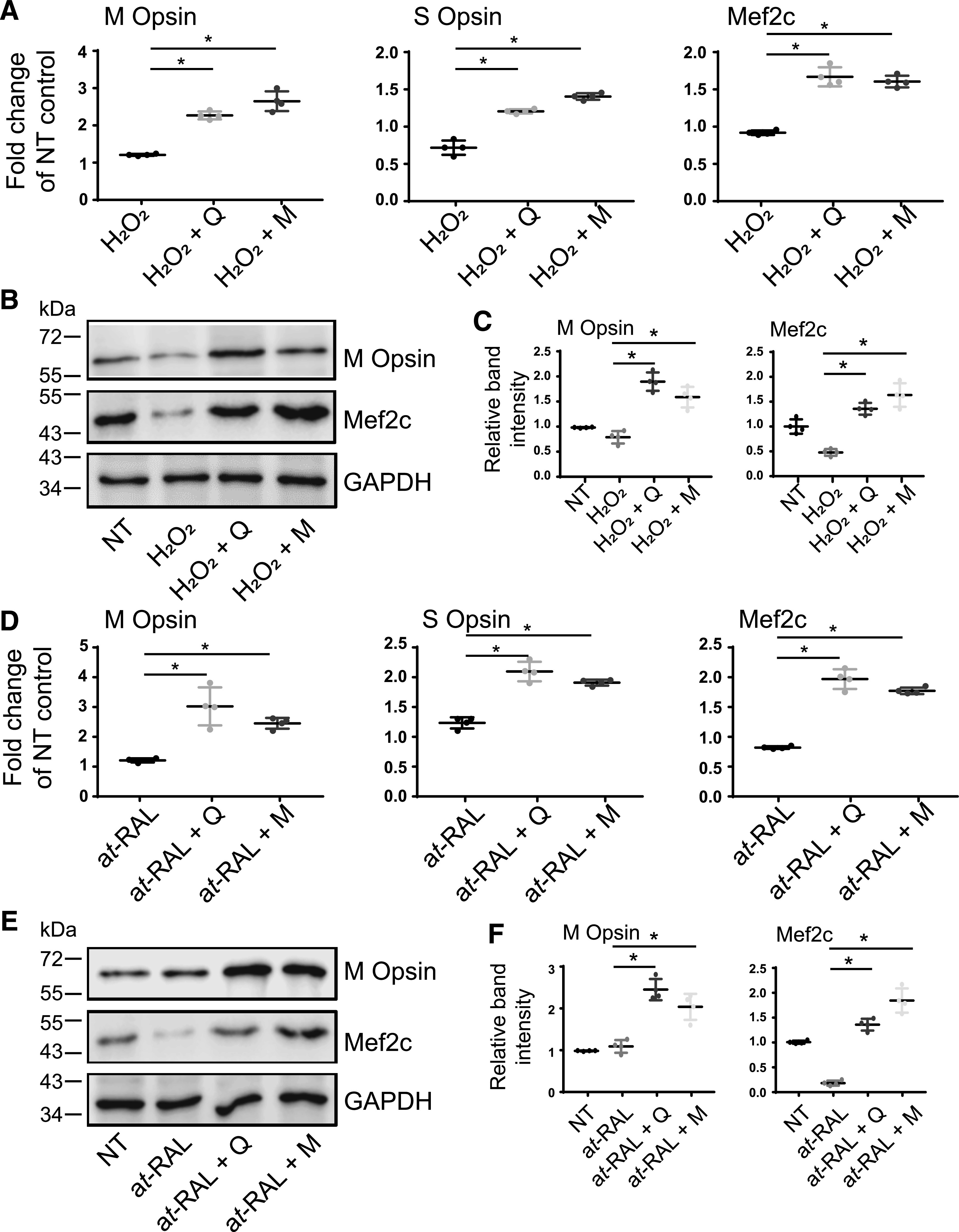
The effect of flavonoids on levels of cone photoreceptor’s specific markers in 661W cells exposed to oxidative stress or all-trans-retinal (at-RAL). 661W cells pretreated with 100 µM quercetin or myricetin for 16 hours were then treated either with H2O2 (250 µM) (A–C) or all-trans-retinal (15 µM) (D–F) for 24 hours. (A and D) RT-qPCR was performed to determine the mRNA expression levels of cone-specific genes, such as M and S cone opsins, and Mef2c, their expression regulator. Relative fold change of these genes’ expression was normalized to the expression of GAPDH. The mean of data from three independent experiments is shown. The expressions of cone opsins and Mef2c were upregulated in cells treated with flavonoids. The statistically significant changes in the expression of the specific gene (M and S cone opsins or Mef2c) compared with stressor (H2O2 or at-RAL)-treated cells are indicated with (*). Error bars indicate S.D. Statistical analysis was performed with the one-way ANOVA and Dunnett’s post hoc tests. (B and E) Immunoblot analysis examining changes in the protein expression of M cone opsin and Mef2c in response to stress and flavonoid treatment. (C and F) Quantification of M cone opsin and Mef2c expression levels. Protein bands were quantified using densitometry analysis with ImageJ software. The mean of data from three independent experiments is shown. The statistically different changes in the expression level of the specific protein (M cone opsin or Mef2c) compared between stressor (H2O2 or at-RAL)-treated cells and flavonoid-treated stressed cells are indicated with asterisk (*). Error bars indicate S.D. Statistical analysis was performed with the one-way ANOVA and Dunnett’s post hoc tests. D, treated with DMSO vehicle; M, treated with myricetin; NT, nontreated; Q, treated with quercetin.