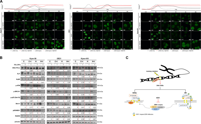
Fig. 5. Effect of IOX1 inhibitor in DNA damage repair after 2 Gy irradiation in ESCC cell lines.
a Representative pictures of nuclear γ-H2AX staining of 2 Gy irradiated ESCC cells treated with 50 µM IOX1 (0–24 h) under 50 µM CoCl2 and hypoxia (0.5–1% O2) conditions. All pictures were taken from Olympus IX51 microscope at ×400 magnification (scale bar 20 μm). ImageJ software (version 1.6.1, from National Institutes of Health) was used for IF quantification and represents a fold change between 2 Gy irradiated cells and non-irradiated control. IF, fluorescence intensity. b Representative images of total XLF (39 kDa), DNA-PKcs (450 kDa), Ku80 (86 kDa), γ-ATM (350 kDa), γ-BRCA1 (220 kDa), γ-p53 (53 kDa), Mre11 (81 kDa), NBS1(95 kDa), RAD50 (153 kDa) protein levels in ESCC cells treated with 50 µM IOX1 under 50 µM CoCl2 and hypoxia (0.5-1% O2) conditions before and after 24 h of 2 Gy irradiation. Fold change values of 2 Gy/0 Gy IOX1 were compared with 2 Gy/0 Gy hypoxic conditions alone. β-Actin (42 kDa) was used as loading control; C - 50 µM CoCl2; C + I - 50 µM CoCl2 + 50 µM IOX1; H – hypoxia; H + I – hypoxia + 50 µM CoCl2. c Schematic representation of DNA damage repair network. DSB, double-strand breaks; DDR, DNA damage repair; HR, homologous recombination; NHEJ, non-homologous end-joining.

