
Fig. 1.
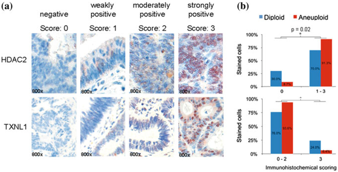
a HDAC2 and TXNL1 immunohistochemical detection in colorectal cancer specimens based on a tissue microarray. Image examples are given at 800-fold magnification. b Tissue-microarray-based immunohistochemical evaluation of HDAC2 and TXNL1 comparing diploid versus aneuploid colorectal carcinoma specimens. Immunoreactivity was scored with “0” showing no positivity, “1” presenting up to 20 % immunopositive cells, “2” up to 50 %, and “3” above 50 % stained cells. Bar plots of the TMA analysis confirmed HDAC2 and TXNL1 as significantly (asterisk) differentially expressed proteins between diploid and aneuploid tumors. Figure adapted from Gemoll et al. (2011)