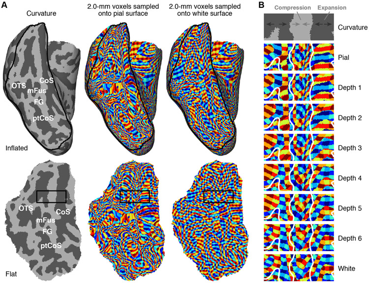
Figure 4. Curvature causes depth-dependent distortion.
A, ‘Surface voxels’ technique. We use nearest-neighbor interpolation to sample isotropic 2.0-mm voxels onto the cortical surfaces for an example subject (S2), and use distinct colors to indicate distinct voxels (see Methods). The top row shows results on the inflated right hemisphere; the bottom row shows results on a flattened section of ventral temporal cortex (indicated by the black outline in the top row). Color patches are relatively isotropic when sampling onto the white-matter surface but are substantially distorted when sampling onto the pial surface. B, Detailed view of rectangular region outlined in panel A. Progressing from inner to outer depths, gyri undergo compression (voxels appear to shrink), while sulci undergo expansion (voxels appear to enlarge).

