Figure 5.
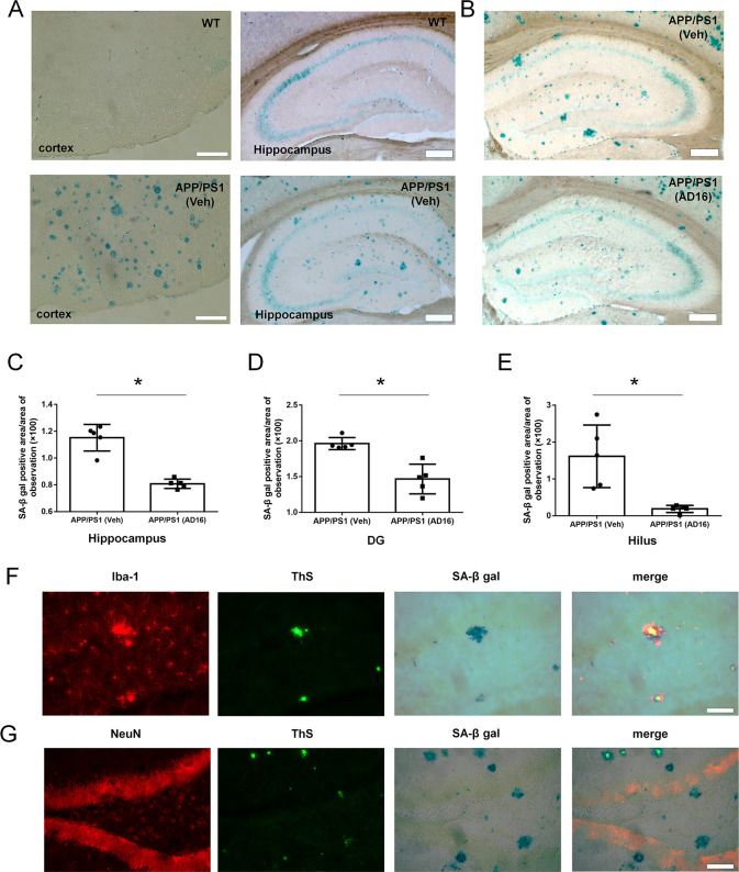
AD16 administration reduces cell aging in the dentate gyrus. Nine-month-old male APP/PS1 mice were either orally dosed with vehicle or AD16 for 3 months. The SA-β-gal activity was increased in the brain tissue of APP/PS1 mice compared to WT mice both in the cortex and hippocampus (A). Representative staining of SA-β-gal in the hippocampus of the APP/PS1 group and AD16 treated group (B). Quantification of the area of SA-β-gal positive cells in the total hippocampus (C), dentate gyrus (DG) of the hippocampus (D), and hilus of the DG part (E) in the brain of AD mice receiving AD16 or vehicle (n = 5 per group). SA-β-gal positive cells were identified through immunostaining with the Iba-1 antibody (F) or NeuN antibody (G). Scale bar: 200 μm. Statistical comparisons were performed by unpaired Student’s t test. Data are presented as means ± SD; *p < 0.05.

