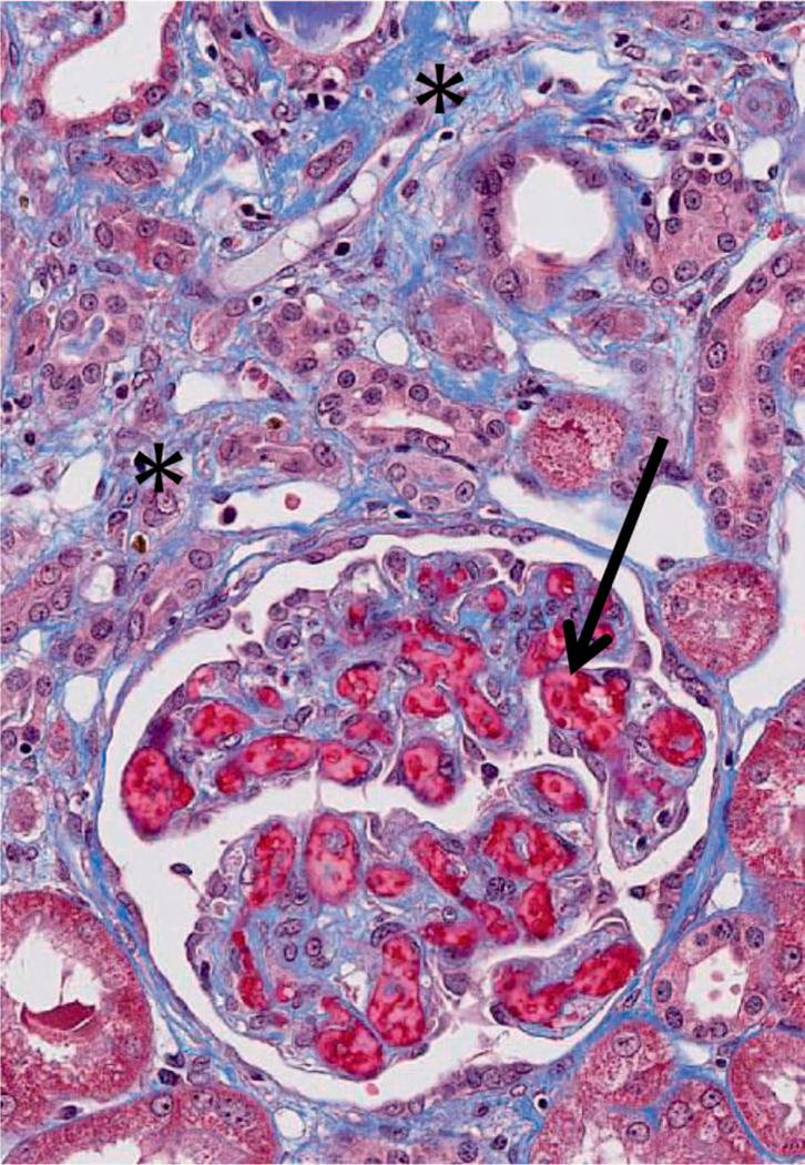
FIG. 9.
Photomicrograph of chronic kidney injury in this NHP model. There is accumulating blue-staining collagenous fibrosis (*), which separates the tubules from each other. There is glomerular capillary thrombosis (arrow). This animal had undergone 11 Gy PBI/BM5 177 days before. Masson trichrome stain, 400×.

