Figure 2.

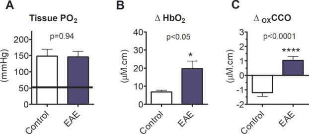
Oxygen administration overcomes spinal hypoxia and improves mitochondrial function. (A) Shows spinal cord tissue PO2 measured by fiber‐optic probe following administration of high oxygen concentrations. The grey shaded area shows the normal range obtained in IFA controls breathing room air. (B and C) These panels respectively show changes derived from trans‐spinal broadband near‐infrared spectroscopy in oxyhemoglobin and oxidation of cytochrome C oxidase. Data presented as mean ± SEM. *p < 0.05, ****p < 0.0001; unpaired t‐test (A: EAE: n = 8; IFA: n = 7; B and C: EAE: n = 11; IFA: n = 9). EAE = experimental autoimmune encephalomyelitis; HbO2 = oxygenated hemoglobin; IFA = incomplete Freund’s adjuvant; oxCCO = oxidation status of cytochrome C oxidase; PO2 = partial pressure of oxygen. [Color figure can be viewed at www.annalsofneurology.org]
