
Fig. 3.
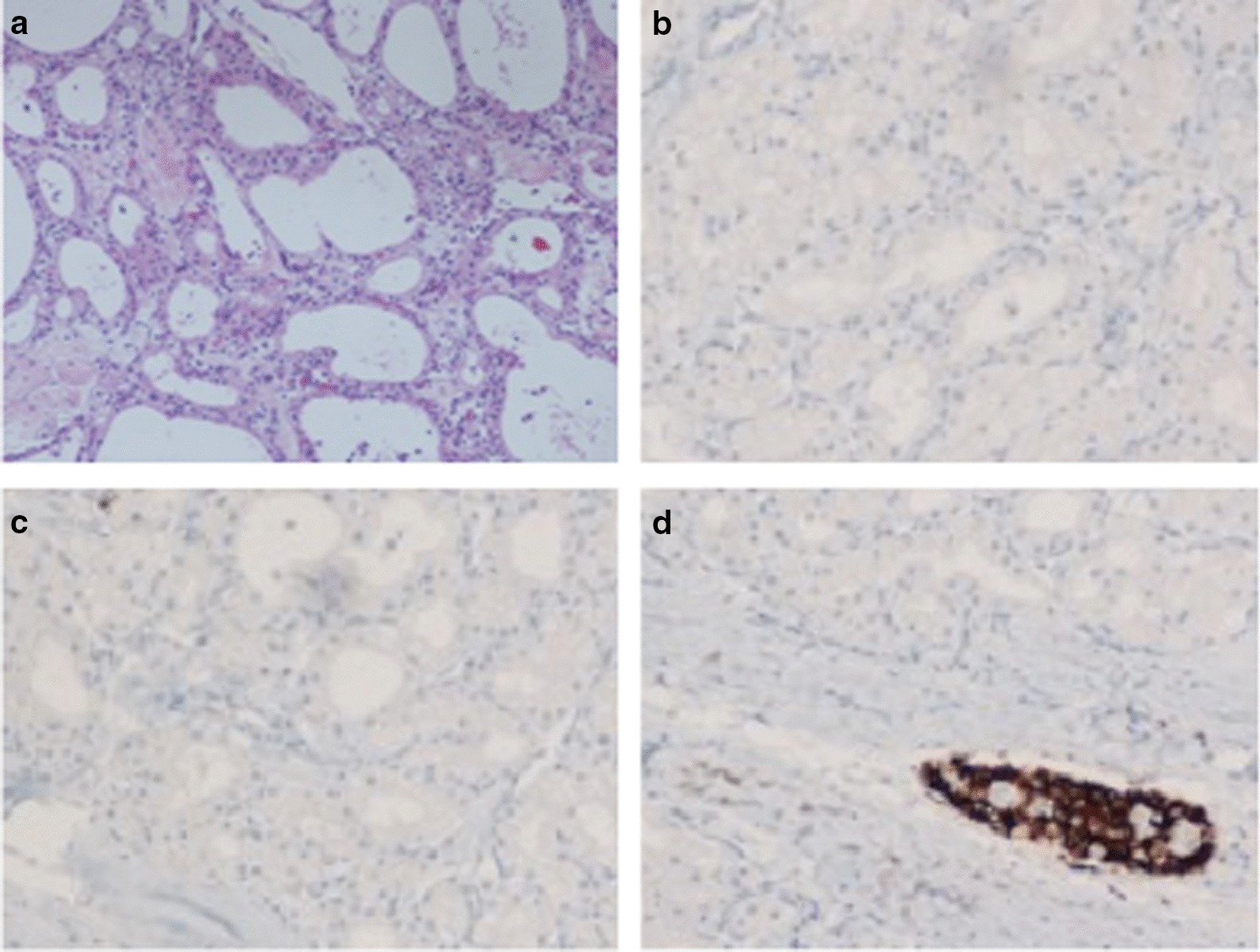
Histology of the specimen: a the cysts were lined by a single layer of uniform, cuboidal, epithelial cells with clear cytoplasm (due to abundant glycogen) and central oval hyperchromatic nuclei with inconspicuous nucleoli; the central scar and radiating septa was composed of hyalinised collagen. Hematoxylin and Eosin staining (magnification 10×); b–d Immuno-histochemical analysis for Synaptophisin (b), Vimentin (c) and Chromogranin (d), in which, only Langerhans’ islet (Red arrow), and not the cystic lesion (Blue arrows), showed positivity for chromogranin A