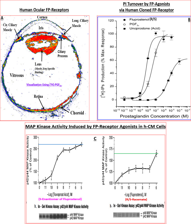
Figure 8.
(A) Autoradiographically visualized FP-receptors in a section of human eye in vitro are shown (total binding of [3H]-PGF2α). The black/white radioautograph was pseudocolor coded to illustrate the relative density of the FP-receptors. Red indicates the highest density, followed by orange, yellow, green, and blue. (B) PI turnover and accumulation of intracellular [3H]-IPs following stimulation by fluprostenol (R/S; racemate), PGF2α, and unoprostone free acid in HEK-943 cells expressing human cloned ciliary body FP-receptor. Reproduced with permission from ref (164). Copyright 2002 Mary Ann Liebert Publishing Inc. (C) MAP kinase activity stimulated by travoprost acid (S-enantiomer of fluprostenol) and fluprostenol (R/S; racemate) in isolated and cultivated hCM cells. Reproduced with permission from ref (162). Copyright 2003 Mary Ann Liebert Publishing Inc.

