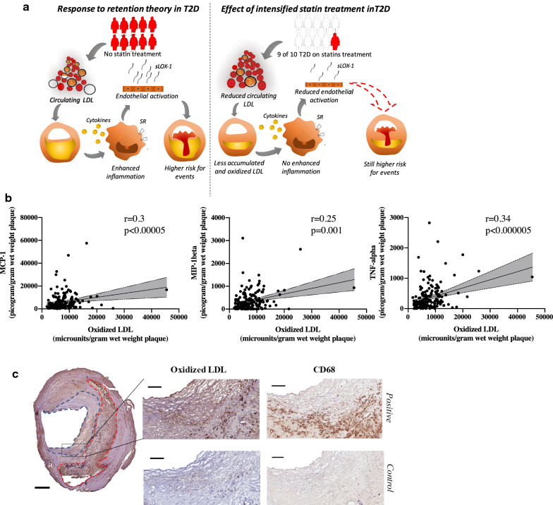
Fig. 1.
a Graphical abstract visualizing the response to retention theory for lipoprotein associated plaque formation and how intensified statin treatment in T2D has affected plaque composition. Individuals marked in red symbolise patients without statin treatment and individuals marked in white symbolise patients receiving statin treatment. LDL, Low density lipoproteins. sLOX-1, soluble LOX-1. SR, scavenger receptors. b Plaque levels of oxidized LDL (oxLDL) correlated to plaque levels of the cytokines monocyte chemoattractant protein-1 (MCP-1), macrophage inflammatory protein-1ß (MIP-1ß), and tumour necrosis factor-α (TNF-α). c Plaque oxLDL was commonly located in the fibrous cap and the core areas and co-localised with CD68 (both stained dark brown). Scale bars 800 µm and in the magnified area 200 µm. Fibrous cap marked in blue dotted line and core in red dotted line

