FIGURE 2.
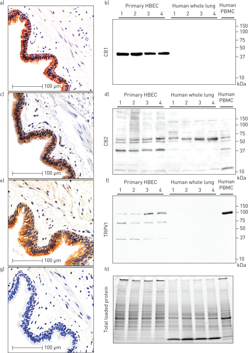
In situ and in vitro validation of CB1, CB2 and TRPV1 protein expression in human airway epithelial cells. Serial sections from a single patient donor that is representative of n=10, for immunohistochemistry of a) CB1, c) CB2 and e) TRPV1 with g) negative control. Immunoblots on primary human airway epithelial cells cultured in vitro: b) CB1, d) CB2, and f) TRPV1 with h) total protein loading control (n=4 airway epithelial cells (HBEC), n=4 whole-lung samples, n=1 peripheral blood mononuclear cells (PBMCs)). Molecular weights (in kilodaltons) are denoted on y-axis of immunoblots.

