
Figure 7. Integrative model of protein degradation and homeostasis in Mycoplasma pneumoniae .
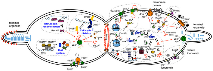
Schematic view of a M. pneumoniae cell during cell division. The right cell compartment shows how protein homeostasis and quality control occur in a minimal cell organism. Protein folding processes with the main chaperones involved are shown: MPN021 (DnaJ), MPN120 (GrpE), MPN331 (Tig), MPN434 (DnaK), MPN531 (ClpB), MPN573 (GroEL), and MPN574 (GroES). Translocation of proteins across the cell membrane through the Sec‐translocation pathway is also shown, including the role of FtsH in regulating its proper assembly. Components of the Sec pathway are MPN184 (SecY), MPN068 (SecE), and MPN396 (SecD). Secreted and membrane‐associated proteins containing signal peptides are delivered to the membrane by MPN210 (SecA) or co‐translationally through the signal recognition particle MPN061 (SRP). All intracellular proteases and peptidases encoded in M. pneumoniae are also represented with their main functions. These include MPN671 (FtsH) and MPN332 (Lon) ATP‐dependent proteases, and peptidases involved in peptide degradation such as MPN022 (Pip), MPN197 (PepF), MPN470 (PepP), MPN572 (PepA), and in protein processing and maturation, such as MPN186 (Map), MPN326 (Prp), and MPN293 (Lsp). Regulation of specific protease and chaperone genes through the MPN124 (HrcA) regulatory system is illustrated. Also the presence of a putative SPase‐I like protein (not yet identified) and the Opp transport system is shown. The left cell compartment shows the major regulatory functions of Lon‐ and FtsH‐controlling specific cellular pathways. Finally, essentiality of the genes involved in this model is shown as E, essential; F, fitness; NE, non‐essential.