Figure 6.

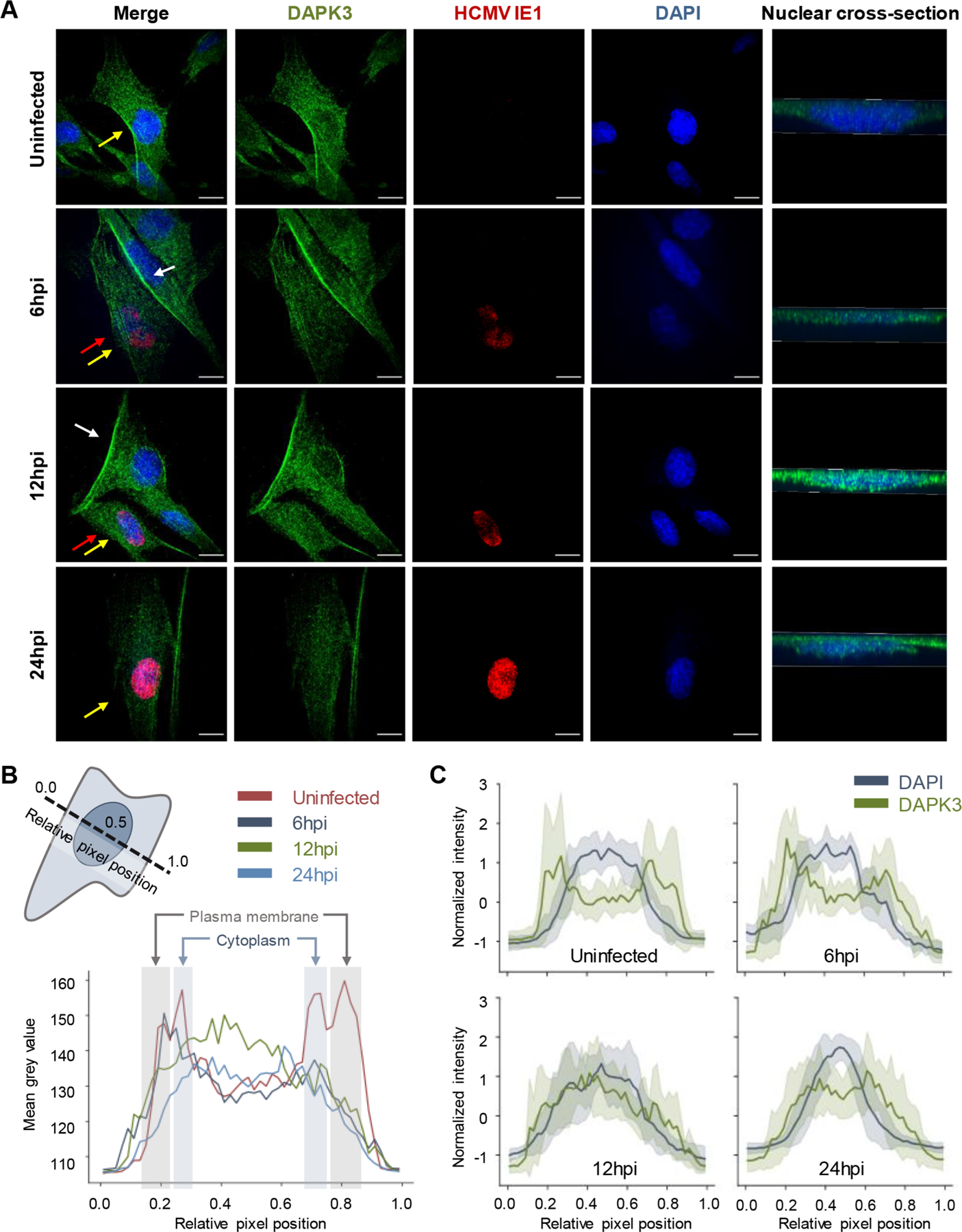
HCMV stimulates DAPK3 translocation between multiple subcellular compartments. (A) Immunofluorescence microscopy images (maximum projections) of DAPK3 distributions in uninfected and HCMV-infected cells early during HCMV infection (6, 12, and 24 hpi). The immediate-early HCMV protein IE1 is provided as marker of infected cells. Yellow arrows denote the nucleus that is cross-sectioned in the right-most panel. For emphasis, white arrows highlight plasma membrane accumulations of DAPK3 in uninfected cells, while red arrows point to infected cells that have lost this phenotype. (B) Line scan analysis of DAPK3 distributions (shaded to highlight the plasma membrane and cytoplasm, with the nucleus between the cytoplasm shadings) in uninfected and infected cells at 6, 12, and 24 hpi. The schematic in the upper-left corner is a representation of the orientation of the line scans relative to the cell body. (C) Overlay of the distribution of DAPK3 relative to the nucleus in uninfected and infected cells at 6, 12, and 24 hpi. Solid lines and shading represent the mean and standard deviation, respectively, across line scans from all cells analyzed at a given condition.
