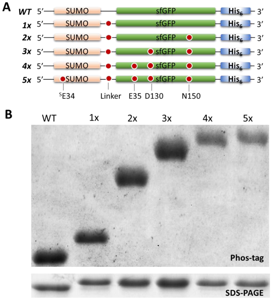
Figure 3.
Incorporation of up to five pSer moieties with pSer-3.1G system. (A) SUMO-sfGFP constructs expressed in pSer-3.1G cells containing the spectrum of zero to five TAG sites. (B) Coomassie stained SDS-PAGE (bottom) and Phos-tag (top) gels showing purified unphosphorylated, singly, doubly, triply, quadruply, and quintuply phosphorylated SUMO-sfGFP. Smaller quantities of protein 4x and 5x pSer proteins were loaded to accentuate the pSer-dependent mobility shift. A Phos-tag gel with higher quantities of protein loaded to evaluate presence of trace quantities of substoichiometrically phosphorylated protein populations is shown in Supporting Figure 11.

