Figure S2.
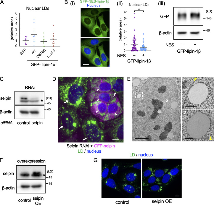
Effects of lipin-1 expression and seipin knockdown and overexpression on nuclear LDs. (A) The effect of wild-type and mutant lipin-1α expression on nuclear LDs. U2OS depleted of lipin-1 was transfected with siRNA-resistant GFP–lipin-1α cDNAs and cultured with OA for 1 d and with OA and 0.25 µM Torin1 (OA/Torin1) for another 8 h. The number of nuclei counted: 9 (GFP), 9 (WT), 10 (D678E), 13 (LxxFF). A representative result of two independent experiments. (B) Comparison of lipin-1β and NES-tagged lipin-1β. (Bi) Distribution of GFP–NES–lipin-1β (green). Nucleus, blue. Scale bar, 10 µm. (Bii) The effect of GFP-NES-lipin-1β expression on nuclear LDs. U2OS depleted of lipin-1 was transfected with either GFP–lipin-1β or GFP–NES–lipin-1β cDNA and cultured with OA for 1 d and with OA/Torin1 for another 8 h. The number of nuclei counted: 54 (lipin-1) or 44 (NES–lipin-1β). Pooled data from three independent experiments. Mann–Whitney test; *, P < 0.05. (Biii) The expression level of GFP–lipin-1β and GFP–NES–lipin-1β (Western blotting). (C) Western blotting of seipin showing U2OS transfected with either control or seipin siRNA. Asterisk indicates a nonspecific band. (D) The effect of seipin re-expression in U2OS treated with seipin siRNA. U2OS after seipin knockdown was transfected with siRNA-resistant GFP-seipin cDNA and cultured with OA for 1 d. Cells expressing GFP-seipin (magenta; arrowheads) show fewer nuclear LDs than those not expressing it (arrows). LD, green; nucleus, blue. Scale bar, 10 µm. (E) EM of U2OS transfected with seipin siRNA and cultured with OA for 1 d. Arrowheads indicate the nuclear envelope. Scale bars, 5 µm; 1 µm (magnified photos). (F) Western blotting of seipin showing control U2OS without cDNA transfection and U2OS transfected with nontagged seipin cDNA. Asterisk indicates a nonspecific band. OE, overexpression. (G) Stable overexpression of seipin decreases nuclear LDs. Cells were cultured with OA for 1 d and OA and 0.25 µM Torin1 for another 1 d. LD, green; nucleus, blue. Scale bars, 10 µm.

