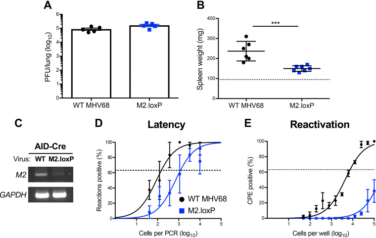
FIG 4.
M2 deletion in AID-expressing cells impairs MHV68 latency and reactivation. AID-Cre mice were infected IN with 1,000 PFU of the indicated viruses. (A) Mice were sacrificed on day 7 postinfection, and viral titers in lung homogenates were determined by plaque assay. (B to E) Mice were sacrificed on day 16 postinfection. (B) Spleens were harvested and weighed as a measure of splenomegaly. The dashed line indicates the average mass of spleens from mock-infected mice. Each dot represents one mouse. (C) DNA was isolated from infected spleens, and PCR was performed to evaluate the integrity of the M2 locus. Cellular GAPDH serves as an amplification control. (D) Single-cell suspensions of spleen cells were serially diluted, and the frequencies of cells harboring MHV68 genomes were determined using a limiting-dilution PCR analysis. (E) Reactivation frequencies were determined by ex vivo plating of serially diluted cells on an indicator monolayer. Cytopathic effect was scored 2 to 3 weeks postplating. Groups of 3 to 5 mice were pooled for each infection and analysis. Results are means of 2 to 3 independent infections. Error bars represent the standard error of the means. *** denotes P < 0.001 in a two-tailed Student’s t test.

