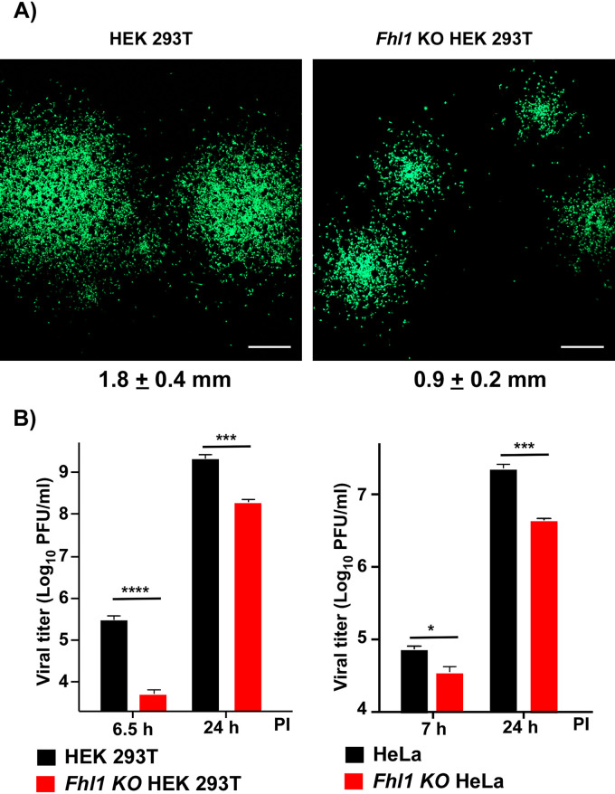
FIG 6.
At low MOI, KO of Fhl1 has a detectable negative effect on CHIKV/GFP titers. (A) Fhl1 KO HEK 293T and parental HEK 293T cells (2 × 10⁶ cells per well) were infected with CHIKV/GFP and incubated for 24 h at 37°C in Avicel-containing media as described in Materials and Methods. Then cells were fixed with 4% paraformaldehyde, and images of GFP-positive foci were acquired on a confocal microscope. Scale bar indicates 0.5 mm. Median diameters of GFP-positive foci and SDs are presented. The difference is statistically significant (P < 0.001). (B) Fhl1 KO and parental HEK 293T and HeLa cells were infected with CHIKV/GFP at an MOI of 0.1 PFU/cell. Viral titers were assessed at 6.5 and 24 h p.i. for HEK 293T and corresponding KO cells and 7 and 24 h p.i. for HeLa and corresponding KO cells. The experiment was repeated 3 times; means and SDs are indicated. Significance of differences was determined by two-way ANOVA Sidak test (*, P < 0.05; ***, P < 0.001; ****, P < 0.0001; n = 3).

